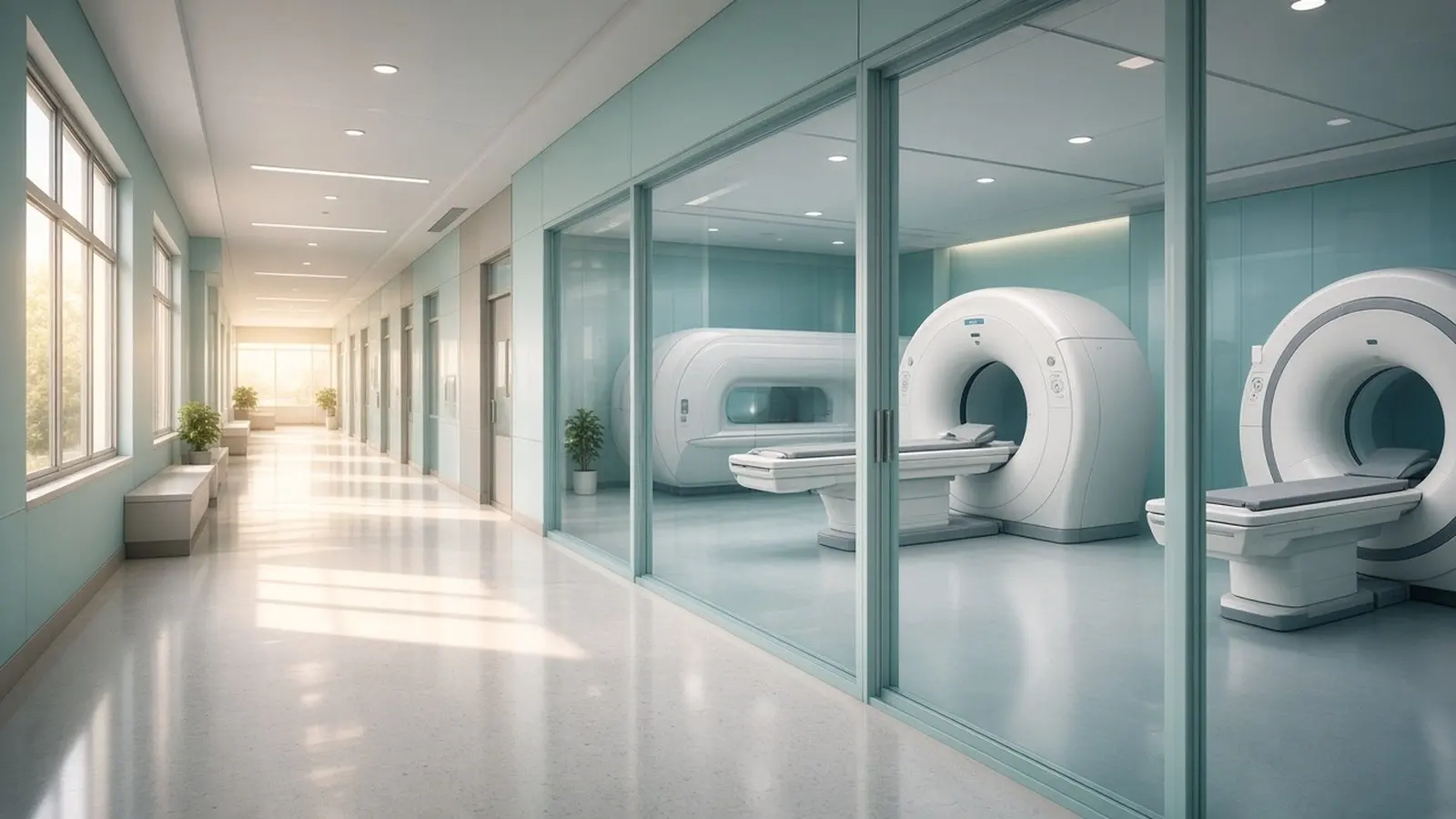
A CT scan uses x-rays to show the shape of organs, bones and blood vessels in a few minutes. An MRI uses a strong magnet and radio waves, with no ionising radiation, to show soft tissue such as the brain, spinal cord, joints and liver in finer detail. A PET scan injects a radioactive tracer, usually a form of glucose, to show where cells are unusually active, which is why oncologists use it, almost always fused with a CT, to find and stage cancer.
How does each scan work?
Each scan builds its picture from a different physical signal. A CT scanner rotates an x-ray tube around you and a computer stacks the thin slices into two- or three-dimensional images; RadiologyInfo notes that CT shows organs, bones, soft tissue and blood vessels in more detail than a plain x-ray. An MRI scanner uses a powerful magnet to line up the protons in the water of your tissues, then knocks them out of line with radiofrequency pulses. NIBIB explains that sensors detect the energy released as the protons realign, and because the timing differs by tissue the machine can tell grey matter from white matter or a tendon from fluid. No x-rays are involved at any stage.
A PET scan images function rather than structure. You receive an injection of a radiotracer, most often F-18 fluorodeoxyglucose (FDG), a molecule similar to glucose. RadiologyInfo explains that cancer cells are more metabolically active and absorb glucose at a higher rate, so they stand out on the image. The tracer gives off gamma rays that a ring of detectors picks up. RadiologyInfo states that combined PET/CT scanners now perform almost all PET scans, so the metabolic map is laid over the CT anatomy, and the combined scan may be more accurate than the two done separately. On an Indian prescription these appear as CT scan, MRI and PET-CT.

What is each scan best for?
CT is the fast all-purpose scan, MRI is the soft-tissue specialist, and PET is the cell-activity scan. RadiologyInfo lists CT as the usual choice for trauma, acute chest or abdominal pain, pulmonary embolism, aortic aneurysm, guiding biopsies, planning surgery or radiotherapy, and detecting cancers of the lung, liver, kidney, ovary and pancreas. CT is less sensitive to movement than MRI, and implanted devices do not prevent it. The FDA states that MRI shows soft tissue better than CT while CT is usually better for bone.
NIBIB states that MRI shows the brain, spinal cord, nerves, muscles, ligaments and tendons more clearly than x-ray or CT, which is why doctors order it for knee and shoulder injuries, and that in the brain it separates white from grey matter and helps diagnose aneurysms and tumours. RadiologyInfo adds that the gallbladder, internal pelvic organs, joints, liver disease, bile duct and pancreas problems, and bowel inflammation in Crohn's disease are often better evaluated with MRI.
PET/CT answers a different question: is this tissue alive and active? RadiologyInfo lists its uses as detecting cancer, showing whether it has spread, judging whether treatment is working, finding a recurrence, assessing heart muscle after a heart attack, and evaluating brain tumours, memory disorders and seizures. The trade-off, per RadiologyInfo, is that PET image resolution is lower than CT or MRI, which is why the two are fused.
How much radiation does each scan give?
MRI gives no ionising radiation, a CT gives roughly one to ten millisieverts, and a whole-body PET/CT gives the most of the three. RadiologyInfo publishes typical effective doses for an average adult and compares each with natural background radiation, which it puts at about 3 mSv per year.
| Scan | Typical effective dose | Equal to natural background of |
|---|---|---|
| Chest x-ray | 0.1 mSv | 10 days |
| CT brain | 1.6 mSv | 7 months |
| CT chest | 6.1 mSv | 2 years |
| CT abdomen and pelvis | 7.7 mSv | 2.6 years |
| CT abdomen and pelvis, with and without contrast | 15.4 mSv | 5.1 years |
| PET/CT, whole-body protocol | 22.7 mSv | 7.6 years |
| MRI | 0 mSv | none |
The FDA puts diagnostic CT doses in the range of 1 to 10 mSv, warns that the estimate for any one procedure can vary by a factor of ten, and states that a 10 mSv CT examination may add approximately 1 chance in 2,000 of fatal cancer to a natural lifetime risk of about 400 in 2,000. The National Cancer Institute gives 4 to 23 mSv for a whole-body PSMA PET/CT depending on protocol, with the PET part alone about 3 mSv, and notes that children are more sensitive to radiation than adults. The NHS says the PET tracer is generally gone within a few hours and that you should avoid prolonged close contact with young children and pregnant people for the period staff tell you.
What are contrast and tracer, and who needs a kidney test first?
Contrast is a dye that makes blood vessels and organs stand out on CT or MRI; a tracer is the radioactive drug that makes a PET image at all. CT uses iodine-based contrast given by mouth, into a vein or rectally. RadiologyInfo states that a serious allergic reaction to it is extremely rare and that departments are equipped to treat one, but that patients with impaired kidney function need special consideration because severe kidney disease raises the risk of worsening kidney function. The NCI says kidney problems can be checked with a blood test beforehand; in India this is the serum creatinine or eGFR that most centres ask for before a contrast CT. Tell the centre about diabetes, thyroid disease, asthma and any previous reaction to contrast, and drink extra fluids afterwards to clear the dye.
MRI uses gadolinium-based contrast. RadiologyInfo states that it is less likely than iodine contrast to cause an allergic reaction, that nephrogenic systemic fibrosis is a rare complication seen in people with serious kidney disease and is exceptionally rare with newer agents, and that the doctor assesses kidney function before injecting it. Trace amounts of gadolinium can remain in organs including the brain; no harm has been identified.
The PET tracer is FDG in most cancer scans. RadiologyInfo says allergic reactions to it are extremely rare and usually mild, and that altered blood sugar or insulin levels can spoil the result, so people with diabetes receive special instructions. The tracer decays on its own and leaves in urine and stool; drinking water speeds this.
How long does each scan take and how do I prepare?
A CT is the quickest at 10 to 20 minutes, an MRI takes 15 minutes to an hour, and a PET/CT visit lasts about two hours. The figures below come from the NHS and RadiologyInfo patient pages.
| CT scan | MRI scan | PET/CT scan | |
|---|---|---|---|
| Preparation | Fast for several hours if contrast is planned; remove metal; declare pregnancy, kidney, thyroid, diabetes, asthma and allergies | Remove all metal and make-up; complete the implant and pregnancy safety form; eat normally | Stop eating 6 hours before, water only; no strenuous exercise for 24 hours; diabetes needs a plan |
| Before the scan | Contrast by drink or injection | Contrast injection if ordered | Tracer injection, then about an hour of quiet rest while it spreads |
| Time on the table | 10 to 20 minutes (NHS) | 15 minutes to 1 hour (NHS) | 30 to 60 minutes (NHS); 20 to 30 minutes of PET plus under 2 minutes of CT (RadiologyInfo) |
| After the scan | Wait up to 30 minutes if you had contrast | No driving for 24 hours if you took a sedative | Drink plenty of water |
| Report | 1 to 2 weeks on the NHS | 1 to 2 weeks on the NHS | 1 to 2 weeks on the NHS |
The NHS advises anyone who is anxious or claustrophobic to say so beforehand; a mild sedative can be prescribed. RadiologyInfo warns that the PET tracer decays quickly, so arriving late may mean the scan is rescheduled.
Who should avoid which scan?
Pregnancy, implanted devices and kidney disease each rule out or change one of the three scans. In pregnancy, RadiologyInfo states that CT is generally not recommended unless medically necessary and that ultrasound or MRI is preferred when it will answer the question; the NCI notes that if CT is unavoidable the dose can be lowered. For MRI, NIBIB states that no effect on the foetus has been demonstrated but that scans are avoided as a precaution, especially in the first trimester, and RadiologyInfo says gadolinium contrast is usually avoided in pregnancy because the risk to the baby is unknown. For PET, the NHS asks you to tell the hospital if you are or might be pregnant or are breastfeeding; staff advise expressing milk before the scan and not breastfeeding for a few hours afterwards, because the tracer passes into milk.
For implants, the issue is MRI. NIBIB states that the magnet is strong enough to fling a wheelchair across the room and that people with implants such as pacemakers, cochlear implants, deep brain stimulators and insulin pumps, should not enter the machine without clearance. RadiologyInfo says most orthopaedic implants pose no risk but asks you to declare every device. CT and PET/CT are not affected.
For kidney disease, RadiologyInfo advises special consideration before iodine contrast and says gadolinium may be withheld; a non-contrast CT or MRI remains possible.
Tell the radiographer before any scan if you are or might be pregnant, are breastfeeding, have a pacemaker or any implant, have kidney disease or diabetes, or have ever reacted to contrast.
In what order are the scans used in cancer care?
Imaging finds and maps a tumour, a biopsy confirms it, PET/CT stages it, and repeat scans track treatment. The NCI states that imaging helps the doctor see whether a tumour is present but that a biopsy is often the only way to tell for sure if you have cancer, so a suspicious scan is normally followed by tissue sampling rather than treatment. The NCI lists CT as the workhorse at every stage: screening, diagnosis, staging, guiding biopsies, planning radiotherapy or surgery, checking response and detecting recurrence. MRI is added where soft-tissue detail decides the plan; RadiologyInfo cites brain, liver, pancreas, bile duct and pelvic tumours, and the NHS notes MRI is used to monitor tumour size during and after cancer treatment.
PET/CT usually enters after diagnosis. RadiologyInfo lists its roles as determining whether cancer has spread, assessing treatment effectiveness, detecting return of disease and evaluating prognosis. An oncologist chooses the order for each cancer type. Ask what each scan will change about the plan before paying for it.
For prices and centres see CT scan cost, MRI scan cost and PET scan cost; in the Tricity, HealthFinder's group company lists published prices at /chandigarh/pet-scan-cost/. This guide is general information from the sources listed below, not medical advice; a registered doctor makes decisions with you.