Take Heed: How many ultrasounds do you have during pregnancy?
The answer to this question is always subjective!
The answer to this question varies from one doctor to another, one patient to another!
Yes,
Depending on your doctor’s & your decision, the ultrasound may take place twice, thrice, or even more within your tenure of pregnancy!
Besides, each pregnancy is different, and ultrasound for each may differ in accordance with – 1) Age, 2) Weight, and 3) Medical History!
Perhaps, there is no ideal count for undertaking an ultrasound before pregnancy. But, you need to know when it usually stands performed and how it has somewhat become a routine test from the moment you conceive!
What organs do they specifically observe during your pregnancy ultrasound? What do doctors look for in an ultrasound during pregnancy?
How do medical professionals proceed with pregnancy ultrasounds? Is there any preparation? If so, what they are? What to avoid during an ultrasound? Are there any dietary limitations?
And,
How beneficial is an ultrasound during pregnancy? What merits do ultrasounds hold?
CContents
How many ultrasounds do you have during pregnancy?
You need to know all of it!
It is because that is when you can determine how many ultrasounds to pursue while you conceive and plan to care for your & your baby’s health. An idea about the various aspects of ultrasound shall let you count this! Of course, it also depends on the amount you can spend on it!
So, in that case, you also need to know – What is the cost of a pregnancy ultrasound. Is there any difference in rate between the different ultrasound techniques? What are they? What are the costs of the latest ultrasound techniques & how different are they from the traditional ones?
Too many questions, bothering your mind, indeed!
But, what if I give you all the answers? If that works, take heed!
First Note –
To know when
During pregnancy, is an ultrasound performed,
You need to fill your knowledge gaps on the stages of pregnancy.
It is because ultrasounds are typically recommended by your doctor following the definite stage you are at!
Medical science classifies the stages of pregnancy into 3 – called the trimesters. So, there exists – A first trimester, a second trimester, and a third trimester!
While a full pregnancy lasts for 40 weeks or so, one trimester involves 12 to 14 weeks from the total.
And,
Often, a fourth stage, the stage of birth, gets associated while it comes to pregnancy ultrasound!
Why so? Let’s find out!
First Stage (or Trimester) –
It is the period starting right at conception and till week 12. In other words, the first trimester entails the moment from when your egg gets fertilized by your male partner’s sperm to the moment the finger & toes grows, stick together, and are identifiable!
It is the first time a pregnancy ultrasound is done to confirm your pregnancy and to estimate how much your fetus has grown. It is, therefore, also known as a fetal ultrasound. It takes place on the 7th or 8th week of pregnancy when the baby’s heart starts breathing, and the embryo is developed rightly in the placenta & amniotic sac and around the length of 1.3 cm.
If the ultrasound result does not show any complication, the next test shall lay undertaken in the second trimester!
And,
If any complications are found, for instance, an ectopic pregnancy where the eggs grow outside the ovaries & in the fallopian tube, another ultrasound may take place during the first trimester, depending on the treatment therapy!
Second Stage (or Trimester) –
In this stage, the fetus is grown with fingers & toes, can swim vigorously, and eyelids are fused. Thereafter, the fetus grows to 14 cm, then 21cm, and finally 33 cm. The completion of the second trimester at the 12th week! The fused eyelids get separated right in this phase, allowing the baby to open & shut its eyes. And it breathes!
The second pregnancy ultrasound takes place right at the beginning of the 12th week when the first trimester ends. And your baby is recognizable on screen. At this time, the ultrasound recommendation comes alongside the recommendation of a blood test to diagnose conditions like trisomy 18 & trisomy 21, the two types of chromosomal abnormalities.
Well, this is the span when your uterus in order to make room for the baby, expands, bulging your belly!
Another round of ultrasound may also take place between the 18th and 22nd week. The gestation period! The ultrasound conducted herein, called mid-trimester ultrasound, shall reveal the position of the fetus, its size, and its surrounding fluids. It may also expose major birth defects like that of the heart or spinal cord & nerves. In fact, other structural defects can even be located, like that of the genitals.
If no defect stands in the way, the next ultrasound takes place in the third trimester!
Third Stage (or Trimester) –
As soon as the baby starts making breathing movements through the lungs, the third trimester takes the stage. This stage lasts till week 40, when the baby is about 51 cm in length, ready to take birth and see the world!
During this phase, two ultrasounds may take place! One in the 28th week and the other between the 32nd to 34th week.
While the former takes place to measure the crown-to-toe length of your baby, the latter look for the preparation of the baby to take birth in a head-down position!
The third-trimester fetal ultrasound involves equipment like an abdominal sensor. And the main objective of this test is to bring out accurate diagnostic information for gynecologists & surgeons to carry out the best possible antenatal care.
Fourth Stage (of Birth) –
A final ultrasound stands done after the baby is born! Of course, we often think ultrasound is unnecessary after the baby is born, but this ultrasound is the most crucial one!
It is called a postpartum ultrasound. After 2 weeks of giving birth, when the postpartum uterus decreases with respect to uterine size & position, this test stands significant! The main goal of this ultrasound is to identify the placenta or product of conception retained within your body. This retained product may be a threat!
An enlarged endometrial cavity is a major consequence of retained pregnancy products or placenta!
On the other hand,
An ultrasound can also serve as a tracing of postpartum endometritis and endometrial infections. The possibility of the latter is 5 to 10 times higher for you if you have a cesarean section!
If you are healthy enough, the ultrasound report shall show normal findings, like intracavitary debris, fluids, & gas!
Second Note –
To learn,
Why & How,
Did the ultrasound technique appear as a routine pregnancy test across the world, You need to know the history of its development.
It is because an ultrasound that gets often attributed for reliability, safety, & efficiency has been modified through time, gradually improving in terms of its application & approach!
No doubt, it is a great contribution to the progress of Gynaecology & Obstetrics!
In the late 1700s (or 18th Century) – Experiments on sound waves began on bats to see how they fly even at night. This experiment lay undertaken by the famous Italian physiologist and priest, Lazzaro Spallanzani (1729 to 1799). The experiment became successful when he was able to determine that a bat, despite, being blind, can fly through space, but not if deaf! Not even if the defect is in only one ear! Therein the idea of sound navigation became clear to science! It was more of a theoretic explanation for bat aviation!
1826 – Another research on sound aviation brought to human history a scientific fact the speed of sound is faster when in the air! It lay done by Jean-Daniel Colladon, a Swiss physician!
1908 – In this year, Austrian Neurologist Karl Dussik attempted to utilize high-frequency sound waves to visualize the size of the brain ventricle & the growth of a tumor. In this experiment, a wand-like instrument, called the transducer, was used. The transducer, at that time, laid on both sides of the patient’s head!
1928 – During this time, ultrasounds lay tested for industrial use. The Soviet Physicist, SY Sokolov, proposed the utility of ultrasound for the detection & screening of flaws in metallic elements!
1949 – This year, George Ludwig, representing the US Naval Military Research Institute, undertook significant research work. It was on the resolution to trace gallstones embedded in the soft tissues. In this experiment, Ludwig applied transmission techniques and subsequently pioneered – How to assess the interactions between animal tissues & high-frequency sound waves?
1956 – The successful use of ultrasound in clinical practices began this year! In Glasgow, An English Physician named Ian Donald tried to measure the diameter of a fetal head with a traducer encompassing one-dimensional amplitude mode.
1958 – High-frequency sound waves lay reused for clinical practices! This time, to scan a female genital tumor!
The 1960s – Tom Brown brought about a revolution in the field of clinical ultrasound. He brought into the picture a two-dimensional scanner. His main objective here was to visualize the density of tissues. Such a development became a turning point for ultrasound application in medical testing! In fact, in 1965, nearly half of the pregnant women in the US took an ultrasound test to see the status of the embryo!
1973 – Another ultrasound approach was being researched at this time! It was a three-dimensional ultrasound, undertaken under the supervision of Tom Brown. The three-dimensional or triplex ultrasound developed more as a matter of necessity outlined by patients! By this time, ultrasound was a popular diagnostic approach, especially for fetal observation & female reproductive health.
The 1980s – The existing, efficient method of screening your digestive & gastrointestinal organs since 1911, endoscopy as we call it, was combined with the techniques of ultrasound to culminate a better imaging process. During this time, the method was developed particularly to improve the screening of tumors in the pancreas & bile ducts, pancreas injury, or obstruction or blockage in the functioning of the pancreas!
At the same, more precisely, in 1986, another innovative ultrasound approach came into action. It was Saline Infusion Sonography. In this method, salt solution, in a small amount, stands used to create proper imaging of the internal structures. Such a technique was primarily meant to identify conditions like endometriosis!
Third Note –
To gather insight on,
What organs a pregnancy ultrasound can diagnose,
You have to understand the main organs associated with pregnancy. They are the main focus of examination herein!
The main organs that your ultrasound team will look at are as follows!
- Uterus & the Uterus Walls.
- Ovaries.
- Fallopian Tube.
- Cervix.
- Placenta.
- Vaginal Opening.
However, in most cases, the organs of the urinary system lay assessed in a pregnancy ultrasound, namely, –
- The Bladder,
- Kidneys,
- Ureter, &
- Urethra.
These are the typical organs your doctor intends to locate & trace abnormalities if any! The type of ultrasound used shall mostly determine the sensitivity to this detection. By this, I mean that some abnormalities may not appear in a traditional ultrasound. In that case, a duplex or Triplex Ultrasound shall lay used!
Fourth Note –
To know,
What exactly your doctor wants to examine about your pregnancy through ultrasound testing,
You have to know,
What condition can an ultrasound diagnose while you bear a child inside you?
If you are pregnant and your doctor asked you to do an ultrasound test, the main reasons behind it can be various. They may be predicting either of the following possibilities!
- Your doctor wants to confirm that baby is growing healthy! She wants to measure the size of the fetus, the heartbeat, or the position it is in!
- Your doctor predicts abnormal conditions like ectopic pregnancy or miscarriage.
- Your doctor wants to see which stage of pregnancy you are at!
- Your doctor wants to know the count of pregnancy, i.e., whether you will have one baby or two or more!
- Your doctor wants an assurance that you shall not have early labor or delivery.
- Your doctor seeks to find out the due date of delivery.
- Your doctor aims to study the placenta that carries nutrients to the fetus.
Fifth Note –
To discover,
The procedure of pregnancy ultrasound,
You must be aware of which ultrasound method you are going to uptake!
Perhaps, there are two different methods of fetal ultrasound, namely, – Transabdominal Ultrasound & Transvaginal Ultrasound!
The process of using sound waves lay the same, but there stands a difference in invasion! Also, lay a difference in the requirement of keeping the bladder full or empty! And the difference in the use of sedatives!
Transabdominal ultrasound involves the creation of imaging through high-frequency sound waves without any invasion of the body. A transvaginal ultrasound, on the contrary, stands minimally invasive! Yes, the transducer, in case of the latter, gets inserted into your body through the vaginal opening.
It is also why one needs sedatives for a transvaginal ultrasound if she cannot bear the pain of insertion, even though the experience of pain is subjective. However, that is not the case for a transabdominal ultrasound. Hence, transabdominal ultrasound can never have side effects. But a transvaginal one can, and that too only because of the application of sedatives.
Apart from these, the rest of the procedure is almost the same. Let us see it all step by step!
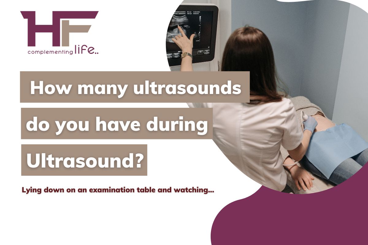
- After you reach the diagnostic center, you shall be taken to the examination hall where various state-of-the-art medical apparatus lay arranged. There exists a large table or bed! Do you see it? The healthcare providers shall ask you to lie down on this table or bed and begin the examination, pull off your dress & expose your abdomen or vagina for the examination!
- The sonographer who conducts the test shall begin his task on the instructions of the radiologist who supervises the ultrasound testing. The individual shall take the transducer and apply a gel-like substance to it. It is the lubricant that serves greatly to – 1) squeeze out the air in between your skin and the transducer when pressed over your skin (for abdominal ultrasound), and 2) reduce painful sensation or discomfort when the transducer is inserted into your vagina (in a vaginal ultrasound).
- For a vaginal or transvaginal ultrasound, an additional step stands by! It is before the sonographer inserts the transducer gets into your vagina! Apart from the lubricant, the sonographer shall use a plastic condom-like sheet to cover the transducer top, further reducing painful sensations, if any!
- Then, the healthcare providers shall request you to bend your knees with your feet at a stir-up position and hold them there for a while. So, the professional can either insert the transducer via your vaginal opening or press & move it over your abdominal skin. It is when sound waves originate from the transducer and travel in different directions within your body.
- If & when the sound waves stumble upon the tissues, walls, organs, veins, or arteries, they reflect the transducer without you hearing any sound or feeling of it passing through! They create faint echoes that turn into electric signals, creating images on the monitor screen.
- When the radiologist aspires to take images from multiple angles, you may be asked to shift to a different position and hold on to it again for a while! But, this step is usually not taken if the test is not a diagnostic one. Instead, just a routine pregnancy test!
- The radiologist shall thereby instruct the sonographer to remove the transducer. And then shall ask you to leave the test lab. Now, you can go to the waiting room, where you get to wash out the additional lubricant and dress up back to how you arrived.
This shall take 15 to 30 minutes at the most! After that, you are free to go back to your normal schedule!
Sixth Note –
To find out,
The entire preparation for an ultrasound,
You have to consult with your doctor because only your doctor can say what is best for you.
However, there are some basic tips from the experts that you may take note of before going for a pregnancy ultrasound!
- First and foremost, you have to check your diet! Usually, doctors recommend a mild one for the day before the test. But, for preparation to start before a week, all that you need to do is avoid spicy food to limit indigestion.
For the day before the test, you can have –
- Some cooked potatoes,
- Breast toast,
- Jelly-O,
- Green Tea, or Coffee,
- Broth,
- Canned fruits & vegetables,
- Fruit juices, and
- Lots of water!
But, there are quite a few meals that you have to avoid like –
- Diary Products,
- All kinds of meats,
- Fresh fruits or vegetables,
- Processed Food.
You need to stop eating at least eight hours before the test and keep your stomach empty until the test is complete! Depending on whether you are uptaking a transvaginal or transabdominal Ultrasound, you shall have limitations on drinking water or fluids. For the former, you need to keep your bladder empty, and thus, cannot drink water on the day of the test until it stands done! However, it is just the opposite for the latter.
- The second step of preparation shall concern medication intake. Typically you can have your regular medications before an ultrasound. However, there is some restriction on the consumption of medicinal drugs that contain aspirin. Painkillers such as–
- Ibuprofen,
- Aspergum,
- Trigesic,
- COPE tablets,
- Anacin,
- Alka-Sheltzer,
- Bufferin, etc., need to be avoided!
- The third step covers the do(s) and doesn’t (s) of what to wear. Medical professionals recommend loose & comfortable attire for an ultrasound. Something that you can slip off easily! At the same time, they ask you to wear any metallic accessories as the presence of metal may tamper with the accuracy of the test result by interrupting the circulation of sound waves.
- The fourth step of pregnancy ultrasound preparation is not mandatory and is only applicable to a transvaginal or vaginal ultrasound. It is the task of removing your pubic hairs if you are uncomfortable exposing your vagina to them!
- The fifth step is to keep all medical documents with you, like your pregnancy reports, your list of medications, your doctor’s prescription, and other test reports. This may be necessary if any documents are asked for at the center reception!
- The final step is to read and sign the consent form before entering the test lab. You may opt to not take sedatives, and that would be a bit of advice from all medical experts as they may bring side effects like –
- Nausea,
- Fatigue,
- Dizziness,
- Sore throat,
- Rashes & redness on the skin near the area of intervention,
- Acne,
- Skin infections, & so forth!
Seventh Note –
To see,
How an ultrasound is beneficial during pregnancy or what as a whole grounds the merit of the process of an ultrasound,
You have to look for the unique features it holds!
No doubt, ultrasound is considered the most significant method of imaging tests and stands out over all other tests!
Are you wondering why so?
It is because –
- An ultrasound is a non-invasive or minimally-invasive process and therefore comes with no pain as such! No pain means No problem! So, you can go back home on the very day of the test. In other words, the test is conducted on an outpatient basis!
- The ultrasound method does not expose you to ionizing radiation and hence is the safest modality to see the fetus. As a result, you and your baby can be free from threats like cell damage or cancer.
- It is a quick process. And you can pursue it any day, any time, without hampering your daily work! You do not have any after-effects. Hence, you also can be on your terms all the time before & after the test!
- This test does not bear any magnetic resonance and thus can be performed on any woman, irrespective of whether you have any implants or not!
- It is cost-effective and can come with a pocket-friendly budget, precisely, around Rs. 500 to Rs. 1500 depending on the type, while you can take care of your pregnancy to the core!
Wrap Up!
Thank you for your time. I hope you have a happy pregnancy!
For more information, stay connected to www.healthfinder.in!









Comments