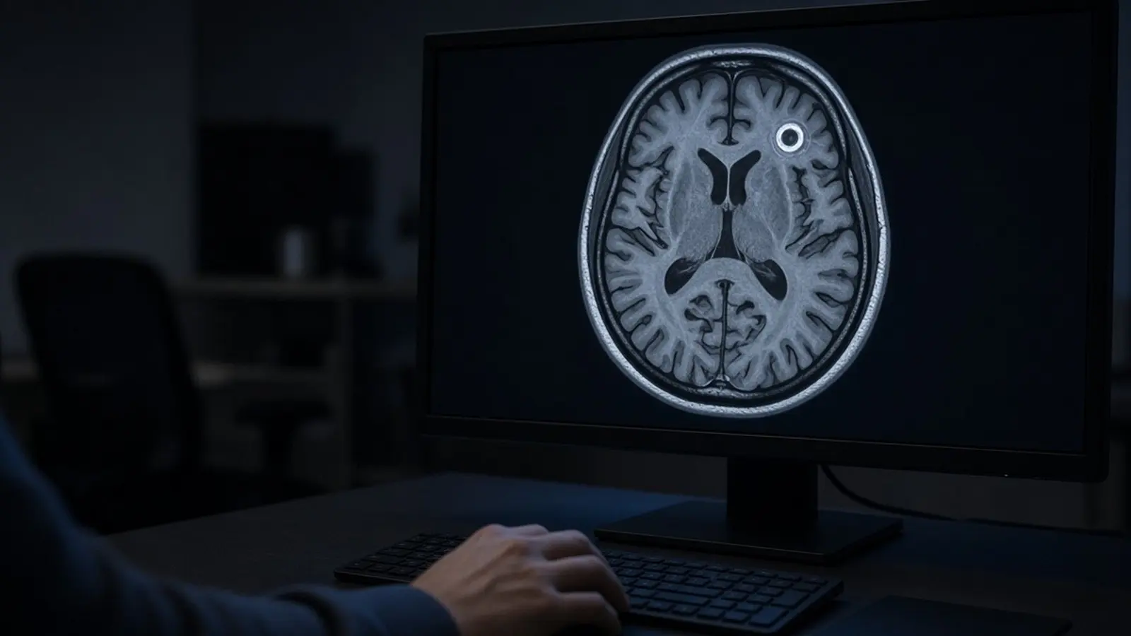
A brain abscess is a pus-filled swelling inside the brain, usually caused by bacteria or fungi that arrive from an infected ear, sinus or tooth, through the blood, or through a head wound. It is a medical emergency: untreated it is almost always fatal, and even with treatment MedlinePlus puts the death rate at about 10% to 30%. Diagnosis is by CT or MRI, and treatment is weeks of intravenous antibiotics, often with surgical drainage.
What is a brain abscess?
A brain abscess is a collection of pus inside the brain tissue, walled off by the body's own defences. The NHS describes it as a pus-filled swelling in the brain that usually forms when bacteria or fungi enter the brain tissue after an infection or a severe head injury. The AIIMS neurosurgery teaching notes define it as a focal infection that begins as a localised area of cerebritis (inflamed brain) and develops into a collection of pus surrounded by a well-vascularised capsule.
It is rare. Cleveland Clinic estimates 1,500 to 2,500 cases a year in the United States, and a 2016 case report in Infectious Diseases in Clinical Practice gives an incidence of 0.4 to 0.9 cases per 100,000 people. The AIIMS notes and a 2022 Indian series both say the condition is more common in developing countries, where untreated ear infections are more frequent.
The danger comes from three things. The pus takes up space inside a closed skull, raising pressure on the brain. The surrounding brain swells, which the NHS says can disrupt blood and oxygen supply. And the abscess can rupture into the fluid-filled spaces of the brain, causing meningitis. Cleveland Clinic names the most common organisms as Staphylococcus and Streptococcus species; the AIIMS notes add that Mycobacterium tuberculosis, fungi and parasites also cause abscesses, which matters in India.

How does infection reach the brain?
Infection reaches the brain by three routes: direct spread from an infected structure next to the brain, through the bloodstream from an infection elsewhere, or straight through a wound. The NHS brain abscess causes page lists the typical sources for each.
| Route | Typical source (NHS) | Notes |
|---|---|---|
| Direct spread from the skull | Persistent middle ear infection (otitis media), mastoiditis, sinusitis, dental abscess | Ear infections tend to seed the temporal lobe and cerebellum |
| Bloodstream | Pneumonia, bronchiectasis, endocarditis, skin infections, pelvic infections, cyanotic (blue) congenital heart disease | Often multiple abscesses |
| Trauma | Skull fracture from a penetrating injury, gunshot or shrapnel wound; rarely neurosurgery | Bacteria enter through the break |
In India the ear is the main culprit. A 2022 retrospective study of 140 brain abscesses at a tertiary hospital in Puducherry, published in Microbiology Insights, found otogenic (ear) infection in 53.6% of cases, and every temporal and parietal lobe abscess in that series was linked to an ear infection. An older Hyderabad series quoted in the AIIMS notes found an ear source in 48% of 50 cases, congenital heart disease in 18% and lung disease in 12%.
The NHS says the source is never found in some cases; the 2016 case report in Infectious Diseases in Clinical Practice puts that figure at about 30%. People with a weakened immune system (HIV, chemotherapy, transplant medicines) and people with cyanotic heart disease are at higher risk.
What are the stages of a brain abscess?
A brain abscess goes through four stages: early cerebritis, late cerebritis, early capsule and late capsule. The 2016 report in Infectious Diseases in Clinical Practice describes the first two with day ranges. Early cerebritis occurs from days 1 to 3: a poorly demarcated area of inflammation with neutrophils, dying tissue and swelling, which on a scan looks like an irregular low-density area that may or may not enhance with contrast. Late cerebritis occurs from days 4 to 9, when macrophages and lymphocytes take over; the area often looks larger on imaging and shows ring enhancement after contrast.
In the capsule stages the body builds a wall of collagen around the necrotic centre, and the scan shows a clear ring enhancing with contrast. The AIIMS teaching notes add that capsule formation depends on the organism, that abscesses seeded through the blood are often poorly capsulated, and that corticosteroids slow capsule formation. Abscesses in newborns, caused by organisms such as Citrobacter and Proteus, are often large with poorly formed capsules.
Why do the stages matter? Early cerebritis has no pus to drain, so antibiotics alone are the treatment. A mature, capsulated abscess has a pocket of pus that a needle can aspirate for culture and relief of pressure. The AIIMS notes also observe that patients improve clinically before the scan changes: improvement on CT is usually seen within one to four weeks, and complete resolution takes one to eleven months, average about three and a half months.
What are the symptoms of a brain abscess?
The most common symptom is a headache, often severe and in one part of the head, that painkillers do not relieve. The NHS and MedlinePlus list the rest: confusion or irritability, fever and chills, nausea and vomiting, stiff neck, seizures, muscle weakness or paralysis on one side, slurred speech or language problems, reduced sensation, and vision changes such as blurred, greyed or double vision from pressure on the optic nerve. Symptoms may appear over several weeks or come on suddenly.
The textbook picture is unreliable. The AIIMS notes say the classical triad of headache, neurological deficit and fever is seen in only 25% of patients. The 2016 report in Infectious Diseases in Clinical Practice says fever is present in less than half of cases, focal neurological signs in about 50%, and seizures in up to 40%. A seizure in an adult who has never had one, especially with a recent ear or sinus infection, should prompt a brain scan.
In babies the NHS adds a bulging soft spot (fontanelle), high-pitched crying, projectile vomiting and stiff limbs.
Call 112 or go to the nearest hospital casualty immediately if someone has a severe headache with slurred speech, weakness or paralysis of an arm, leg or one side of the face, a first-ever seizure, or drowsiness that is getting worse. The NHS treats these as emergencies. Fever with a worsening headache and vomiting needs a doctor the same day.
How is a brain abscess diagnosed?
A brain abscess is diagnosed with a contrast CT or MRI scan of the head, supported by blood tests and, when safe, a needle sample of the pus. The NHS says doctors first assess symptoms, recent infections and immune status, then send blood tests for infection and refer the patient to hospital for a CT scan (a series of X-rays) or an MRI scan (magnetic fields and radio waves). MedlinePlus adds blood cultures, a complete blood count, chest X-ray, EEG if there are seizures, and antibody tests for specific germs.
On a contrast scan a mature abscess shows the typical ring: a dark centre of pus, a bright enhancing capsule and swelling around it. MRI distinguishes the stages better; the AIIMS notes include MRI appearances for early cerebritis, late cerebritis and the capsule phases. See HealthFinder's MRI scan cost and CT scan cost pages for price ranges by city; in an emergency the scan is done in hospital, not booked separately.
Identifying the organism is harder. The NHS describes CT-guided aspiration, where a needle guided by the scan removes pus for testing. MedlinePlus says a needle biopsy is usually performed to identify the cause. Even so, cultures are often negative: the Puducherry series found only 47.14% of samples culture-positive, and the 2016 report notes that blood and spinal fluid cultures identify the organism in only about 25% of cases. A lumbar puncture is not a routine test here; with a mass in the brain it carries risk and is avoided unless meningitis must be excluded.
How is a brain abscess treated?
Treatment is intravenous antibiotics (or antifungals) for weeks, plus drainage when the abscess is large or not shrinking. The NHS says antibiotics often begin before the diagnosis is confirmed, through a drip into a vein. Cleveland Clinic says antibiotics are needed for at least four to eight weeks; the AIIMS notes give six to eight weeks, because the brain takes long to repair and close the abscess space.
When is surgery needed? The sources agree on the broad rule and differ slightly on the size:
- Medicines alone are preferred when the abscess is small (under 2 cm per MedlinePlus and Cleveland Clinic, under 2.5 cm per the NHS), deep in the brain, multiple, or accompanied by meningitis.
- Aspiration or surgery is used when the abscess is larger than that, pressure in the head is rising, the abscess does not shrink on antibiotics, it contains gas, or it may rupture. The AIIMS notes cite Rosenblum's finding that no abscess over 2.5 cm resolved with antibiotics alone.
Simple aspiration, per the NHS, means drilling a small burr hole in the skull under CT guidance and draining the pus; it takes about an hour. A craniotomy, removing a flap of bone to take out the abscess, takes about three hours and is used when aspiration fails or the abscess returns. The pus goes to the lab to match the antibiotic to the organism. Steroids and diuretics may reduce swelling; anti-seizure medicines are common.
Treatment needs a hospital with a neurosurgery unit; government medical colleges treat brain abscesses at low cost.
What is the outlook after a brain abscess?
Most people survive a treated brain abscess, and outcomes are better the earlier treatment starts. MedlinePlus says that untreated it is almost always fatal and that with treatment the death rate is about 10% to 30%. The AIIMS notes record a fall in mortality from 40 to 60% before CT scanning to 0 to 10% today, crediting earlier diagnosis, better culture techniques for anaerobes and antibiotics that cross into the brain. The 2022 Puducherry series of 140 patients reported no deaths.
Recovery is slow. The NHS says most people stay in hospital for several weeks and then need a further 6 to 12 weeks of rest at home before returning to work or education, and should avoid contact sports with a risk of skull injury. Repeat CT scans confirm the abscess has gone. Rehabilitation with a physiotherapist or speech therapist helps with weakness or speech problems.
The complications the NHS and MedlinePlus list are:
- Brain damage: mild to moderate damage often improves with time; severe damage is likely to be permanent, and delay in treatment raises the risk.
- Epilepsy: repeated seizures, which may need long-term medicine.
- Meningitis: life-threatening if the abscess ruptures.
- Recurrence, more likely in people with weak immunity or cyanotic heart disease; any return of symptoms needs immediate review.
A neurologist usually manages follow-up for seizures and residual weakness after the neurosurgeon's part is done.
How can a brain abscess be prevented in India?
Prevention means treating the infections that seed the brain, above all chronic ear discharge. Because more than half of Indian cases in the Puducherry series came from the ear, a child or adult with a persistently discharging ear, or pain and swelling behind the ear, needs an ENT specialist rather than repeated courses of drops. The NHS lists persistent middle ear infection, mastoiditis and sinusitis as the direct-spread sources, and a dental abscess among the bloodstream sources, so a tooth abscess should be treated promptly too.
MedlinePlus adds that people with certain heart disorders may be prescribed antibiotics before dental and other procedures, and that treating lung infections such as pneumonia and bronchiectasis lowers the risk. People on immune-suppressing treatment should report any fever with headache early.
Tuberculosis deserves a mention in India. The AIIMS notes describe the tubercular abscess as rare, containing live TB bacilli, and treated mainly with anti-tubercular therapy; the Puducherry series isolated Mycobacterium tuberculosis in one case. A doctor who suspects TB will test the aspirated pus for it.
This guide is general information from the sources listed below, not medical advice; a registered doctor makes decisions with you.