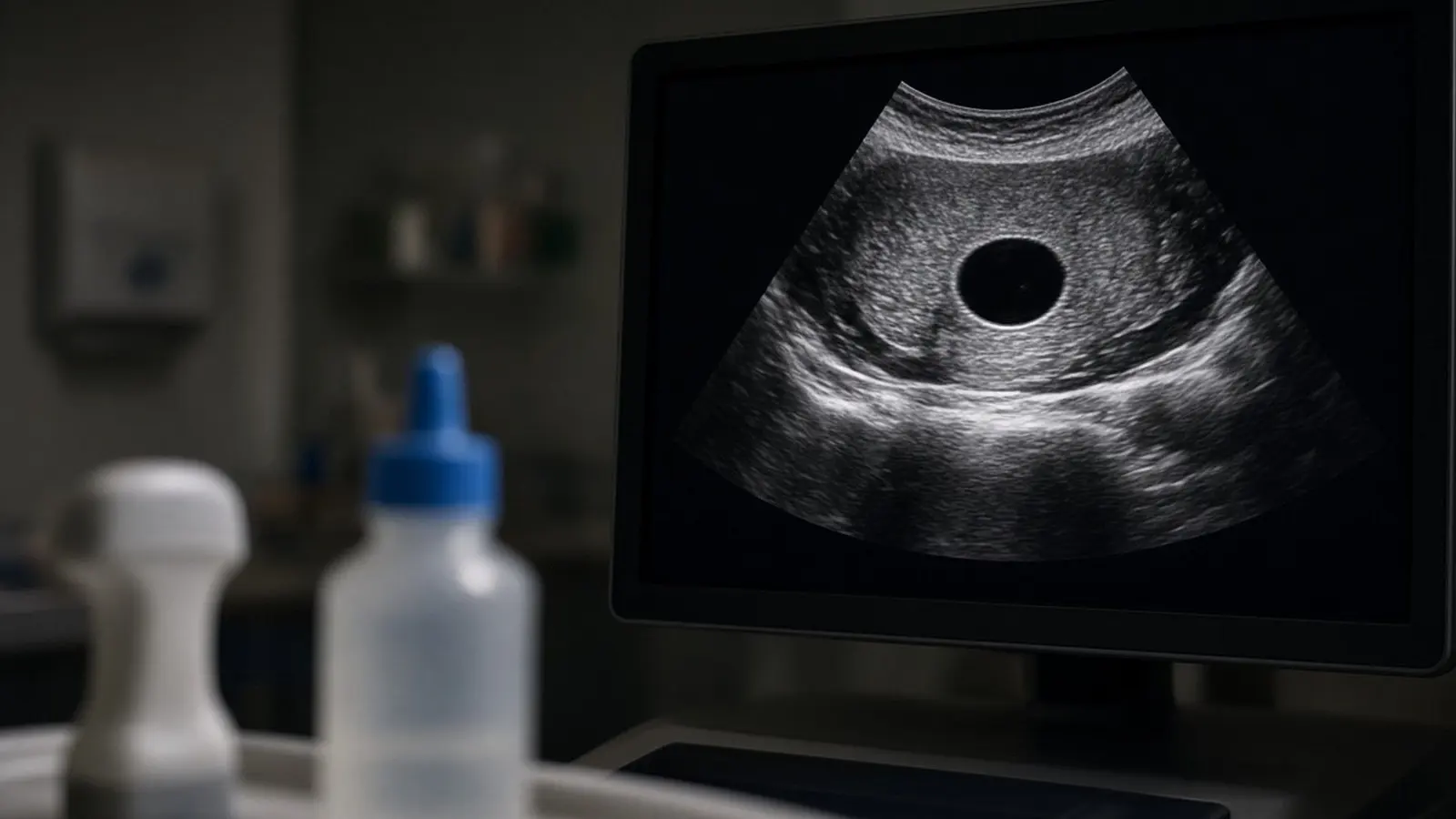
On an ultrasound a simple cyst looks like a black (anechoic), round or oval sac with a thin smooth wall and a brighter band of tissue behind it, which radiologists call posterior acoustic enhancement. Fluid gives no echoes, so it appears black; solid tissue scatters sound and appears grey. A cyst becomes "complex" when it shows internal echoes, thick walls or septa, or a solid part with blood flow, and those are the features that lead to a repeat scan, an MRI or a biopsy.
What do cysts look like on an ultrasound?
A cyst looks like a black, round or oval pocket with a thin, sharp outline and a bright strip behind it. The 2010 Society of Radiologists in Ultrasound consensus statement in Radiology (Levine and colleagues) gives the formal definition for the ovary: "a round or oval anechoic space with smooth thin walls, posterior acoustic enhancement", no solid component or septation, and no internal blood flow on colour Doppler. A 2025 narrative review in Current Medical Imaging (Ceri and colleagues) describes a simple liver cyst in the same words: anechoic, round, well-defined borders, no visible wall and posterior acoustic enhancement. For the kidney, a 2021 review in Medicina (Nicolau and colleagues) defines a simple cyst as fluid with "a thin (≤2 mm) and well-defined wall" and no septa or nodules.
Three words recur on reports:
- Anechoic means black: the fluid returns no echoes.
- Posterior acoustic enhancement means the tissue directly behind the cyst looks brighter than its neighbours.
- Thin-walled or imperceptible wall means the edge is a sharp line, not a thick rim.

The picture is the same whichever organ holds the cyst, so the radiologist describes the shape, the wall, the inside and the size in centimetres, then names the organ. On a printed Indian report you will read lines such as "well-defined anechoic lesion with posterior acoustic enhancement, no internal echoes or septations, suggestive of a simple cyst". That sentence, with the size, is what the rest of this guide explains.
How does ultrasound tell fluid from solid?
Ultrasound tells fluid from solid by what comes back. The probe sends sound into the body and listens for echoes; clear fluid sends almost nothing back, so a cyst is black, while solid tissue scatters the sound and comes back as shades of grey. Cleveland Clinic's breast cyst page puts the practical result simply: a breast ultrasound "will show if the lump is solid or filled with fluid", and fluid-filled cysts are unlikely to be cancerous while hard lumps need further testing.
Radiologists use four checks, all taken from the 2010 consensus and the Medicina review:
| Check | Fluid (simple cyst) | Solid or complex |
|---|---|---|
| Inside (echogenicity) | Anechoic, black | Grey echoes, or a mix of black and grey |
| Behind the lesion | Posterior acoustic enhancement (brighter) | No enhancement, or a shadow |
| Wall | Thin, smooth, 2 mm or less in the kidney | Thick, irregular, or focal thickening of 3 mm or more |
| Colour Doppler | No internal flow | Flow inside a solid part or septum |
Colour Doppler is the decisive check. The consensus statement says "a cyst with a nodule that has internal blood flow has the highest likelihood of being malignant". The Medicina review adds that complex cysts are "usually secondary to the presence of intracystic hemorrhage or infection", which is why old blood in an ovarian cyst or a kidney cyst can look grey without being a tumour.
What is the difference between a simple cyst and a complex cyst?
A simple cyst meets every one of the criteria above; a complex cyst fails at least one. The 2010 consensus lists the features that move a cyst into the worrying group: thick septations of 3 mm or more, focal wall thickening of 3 mm or more, and solid elements with blood flow on Doppler.
| Feature | Simple cyst | Complex cyst |
|---|---|---|
| Interior | Black | Low-level echoes, fishnet pattern, fluid level |
| Septations (internal walls) | None | Thin (under 3 mm) or thick (3 mm or more) |
| Wall | Thin, smooth | Thick, irregular, focal thickening of 3 mm or more |
| Solid part or nodule | None | Present; highest concern with Doppler flow |
| Calcification | None | Present |
| Doppler flow | None inside | Flow in septa, wall or nodule |
Not every complex cyst is dangerous. The consensus describes three common benign patterns in the ovary: a haemorrhagic cyst with a reticular (fishnet) pattern of echoes and no flow, an endometrioma with homogeneous ground-glass echoes and no internal flow, and a dermoid with bright lines and dots and acoustic shadowing. Cleveland Clinic's breast page gives numbers for one organ: simple cysts are always non-cancerous, complicated cysts carry a cancer risk under 2%, and complex cysts with solid parts are cancer in about 14% to 23% of cases, so they are biopsied.
Where are cysts commonly found, and how common are they?
Cysts turn up most often in the ovary, kidney, liver, thyroid and breast, usually by chance.
| Site | How common | Source |
|---|---|---|
| Ovary | "Very common"; most are functional and gone within a few months; a cyst under 3 cm is physiological | NHS; Levine 2010 |
| Kidney | About 1 in 10 people, nearly 1 in 5 over 50; up to half of people over 50 have at least one | NIDDK; Cleveland Clinic |
| Liver | 2 to 5% of ultrasound studies and up to 18% of CT scans; more with age and in women | Ceri 2025 |
| Thyroid | Nodules in 20 to 76% of adults on ultrasound; under 6.5% are cancer; cystic nodules biopsied over 2 cm | Cleveland Clinic |
| Breast | About 7% of women; simple cysts are 90% of breast cysts and always benign | Cleveland Clinic |
The ovary is the special case because cysts there come and go with the cycle. The NHS says functional cysts form as part of the menstrual cycle, are usually harmless and short-lived, and that most cysts disappear within a few months. The NIDDK says simple kidney cysts are "usually harmless" and rarely cause symptoms. Cleveland Clinic says only about 5% of liver cysts are cancerous and that a hydatid (parasitic) cyst "can be fatal without treatment".
If the cyst is in a woman's pelvis the report goes to a gynaecologist; a kidney cyst that needs follow-up goes to a nephrologist or urologist.
Which features on the scan prompt further tests?
Further tests follow when a cyst has a solid part, blood flow, thick walls or septa, a large size, or a patient past the menopause. The 2010 consensus says cysts with a nodule that has internal flow should be considered for surgical evaluation, and that simple ovarian cysts over 7 cm should have an MRI or surgical opinion.
The usual next steps, by organ:
- Ovary. A repeat scan in a few weeks (NHS) to see whether a functional cyst has gone; blood tests for chemicals that can be raised in ovarian cancer; an MRI for a cyst over 7 cm (Levine 2010).
- Kidney. A contrast CT or MRI for any complex cyst, because the Medicina review says "up to 10% of renal cell carcinomas can present as complex cystic masses"; CT and MRI give the Bosniak grade.
- Liver. Ultrasound alone is enough for a simple cyst, per the Ceri review, but septa, wall thickening, nodules or calcification need CT or MRI.
- Thyroid. A fine-needle biopsy, which Cleveland Clinic says is done for cystic nodules larger than 2 cm, or a repeat scan after a few months (RadiologyInfo).
- Breast. Biopsy for a complex cyst (Cleveland Clinic).
See a doctor the same day if a known ovarian cyst gives sudden, severe abdominal pain with vomiting or fever, or if you feel faint with rapid breathing and cold, clammy skin. Cleveland Clinic lists these as signs of a twisted or ruptured cyst. In India call 112 or go to the nearest emergency department.
What do O-RADS, Bosniak and TI-RADS mean on my report?
O-RADS, Bosniak and TI-RADS are scoring systems that turn the features above into a risk category for the ovary, kidney and thyroid.
Bosniak (kidney). The Medicina review (Nicolau 2021) says the classification is built on contrast CT and "is predictive of the risk of malignancy". Bosniak I and II cysts need no further management; IIF cysts need follow-up because of a 5 to 10% risk of malignancy; Bosniak III and IV lesions have about a 50% and 90% probability of renal cell carcinoma and surgery is recommended. A simple cyst on ultrasound is Bosniak I; the grade is not assigned from a plain ultrasound.
O-RADS (ovary). A 2026 review in Frontiers in Oncology (Yu and colleagues) describes the American College of Radiology's ovarian-adnexal system as a 1 to 5 scale grouped into three tiers: benign (O-RADS 1 to 2) managed with conservative follow-up, intermediate risk (O-RADS 3) needing further evaluation or closer surveillance, and high risk (O-RADS 4 to 5) needing surgery or oncology referral.
TI-RADS (thyroid). A 2022 meta-analysis in Cancers (Kang and colleagues) explains the ACR version: five features are scored and summed, giving TR1 benign (0 points), TR2 not suspicious (2), TR3 mildly suspicious (3), TR4 moderately suspicious (4 to 6) and TR5 highly suspicious (7 or more). Biopsy thresholds for TR3, TR4 and TR5 are 2.5 cm, 1.5 cm and 1 cm respectively. A purely cystic nodule sits at the benign end.
What happens after a cyst is found on ultrasound?
After a cyst is found, most people are told to do nothing or to come back for one repeat scan; a minority go on to CT, MRI, biopsy or surgery. The follow-up intervals in the cited sources:
| Site and finding | Follow-up | Source |
|---|---|---|
| Ovary, simple cyst, still having periods, up to 5 cm | None | Levine 2010 |
| Ovary, simple cyst, still having periods, 5 to 7 cm | Yearly ultrasound | Levine 2010 |
| Ovary, simple cyst, after menopause, 1 to 7 cm | Yearly ultrasound at first | Levine 2010 |
| Ovary, any simple cyst over 7 cm | MRI or surgical opinion | Levine 2010 |
| Kidney, simple cyst | None, or periodic ultrasound | NIDDK |
| Kidney, Bosniak IIF | Imaging every six months to two years | Cleveland Clinic |
| Liver, benign cyst under observation | Imaging every three months for a year | Cleveland Clinic |
| Breast, complicated cyst | Return visit in six months | Cleveland Clinic |
The NIDDK describes sclerotherapy for a symptomatic kidney cyst: the cyst is drained under ultrasound guidance and alcohol is injected so it does not refill. Cleveland Clinic says liver cysts over 4 cm count as large and lists needle drainage or removal of the cyst wall for them.
See ultrasound cost in India and MRI scan cost. This guide is general information from the sources listed below, not medical advice; a registered doctor makes decisions with you.