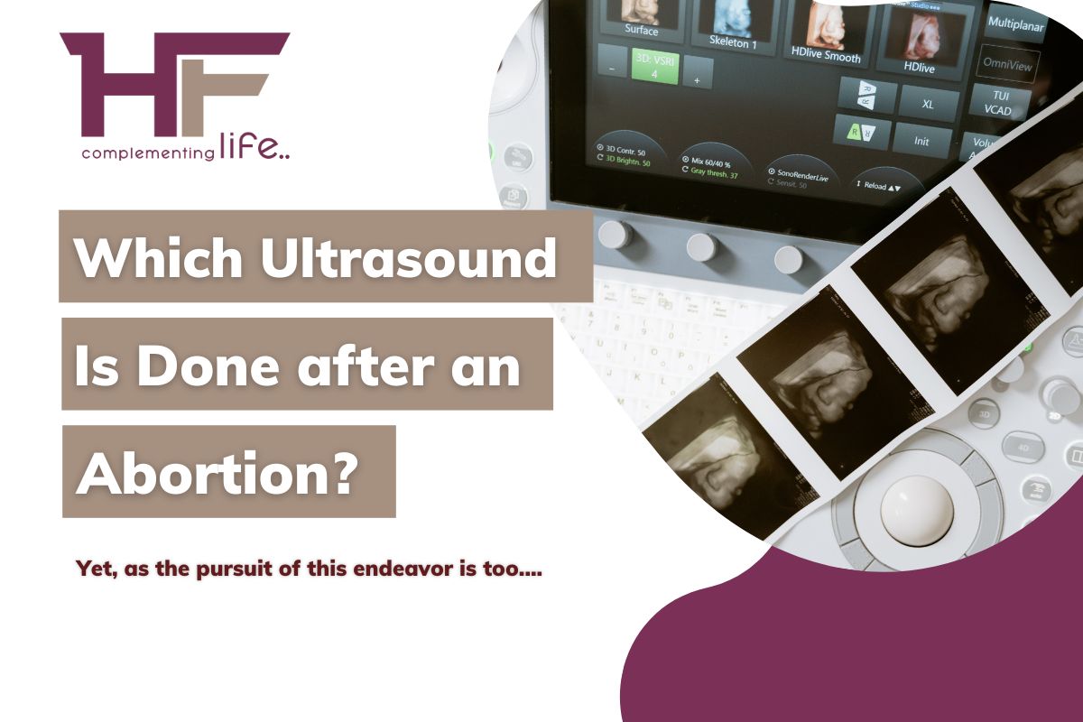
Which Ultrasound is done after an Abortion? No doubt, the development of medical science, precisely in the field of diagnosis, brings before you a versatile diagnostic method called ultrasound. This method is nothing but the use of sound waves in creating images of your internal body, the cells, tissues, organs, and so forth.
CContents
Which Ultrasound is done after an Abortion?
Yet, as the pursuit of this endeavor is too diverse, ultrasound may stand different for examining different health conditions.
And,
This particular difference solely revolves around the area or part of the examination!
For instance, when your doctor predicts and wants to examine a condition like a breast lump, she is likely to use the method of ultrasound called Breast Ultrasound. On the other note, if she wants to find out whether you have stones on your kidney, the technique that she shall execute is the Renal Ultrasound.
Yes,
In a similar fashion, the ultrasound that lay undertaken after an abortion is the one to examine your reproductive organs via your abdominal or vaginal areas, and therefore, regarded as Transabdominal & TransvaginalUltrasound, respectively.
The variation in the practice site is what makes an ultrasound after abortion of two different types, as stated above. Herein, a Transvaginal Ultrasound is more invasive than the Transabdominal one. That is certainly why the requirements of high-level disinfection make the technique pretty cost-prohibitive when compared with the latter Transabdominal sonography!
Wait!
[It is essential to note that the kind of abortion we are talking about now is a medical abortion, not a surgical one. It is because multiple survey records affirm that medical abortion stands more commonly practiced in countries like the US, UK, Australia, Canada, and India.]
For a medical abortion, although the World Health Organization (WHO), the National Abortion Federation (NAF), & the Society of Family Planning (SFP) do not recommend the use of ultrasound on a routine basis, however, most doctors and medical professionals tend to do so. They regard it as a part of the clinical protocols of the institution or practice!
Now,
Many questions are definite to pop up in your mind, and resolving them with accurate insight is our job. That is why we are here today! This article shall help you with a detailed note on your post-abortion ultrasound or ultrasonography.
So, let us start!
What do doctors look for in a post-abortion ultrasound?
Usually, doctors look for a confirmation of the success of abortion via ultrasound testing, especially if you are unsure about it and want valid proof of pedigree to get a sigh of relief.
Yet again, there are other instances in which your doctor may call for a post-abortion ultrasound. Are you wondering what they look for in those instances? Here goes!
Endometrial Thickness & Possibility of Endometrial Cancer in the Near Future
Well, your doctor may seek to look at the endometrial thickness in the first place, and that is one instance in which he or she may urge you to undertake follow-up ultrasonography, particularly after a medical abortion.
It is because the endometrial thickness varies after a medical abortion, turning heterogeneous and complex in its appearance. Such a facet can also sometimes overlap between conditions of successful and unsuccessful pregnancy.
Perhaps, when the endometrial thickness extends beyond 14mm, you are likely to pose serious health threats as dangerous as cancer. Yes, this condition in which the lining of your uterus turns unusually thick due to the existence of excessive cells is called Endometrial Hyperplasia, and it can lead to endometrial cancer.
Multiple studies note that the possibility of cancer development herein stands at around 6.7%, and your doctor does not want you to fall into that percentage. It is precisely why she is insisting on you for a post-abortion ultrasound!
Persistent Gestational Sac & the Future Possibility of Persistent Trophoblastic Diseases
Persistent Gestational Sac is the condition in which even though the viable embryonic tissue is not present, the sac is!
Relax! You don’t need to panic right away, as this condition may occur in less than 1% of the women undergoing medical abortion through the intake of mifepristone and misoprostol.
However, your doctor shall aim to ensure that such a condition does not occur at all, specifically if you take oral misoprostol for the abortion. It is because that can result in a persistent sac with viable gestation in around 9% of women.
Besides, when you undergo an abortion because your pregnancy was molar, i.e., a complex pregnancy where the trophoblast grows abnormally in the placenta, your doctor is skeptical about the remaining of such abnormal tissues in your womb.
If that happens, even in a small proportion, a possibility for these tissues to further develop in the lining of your womb remains. Just like cancer! Medical science refers to this condition as a persistent trophoblastic disease, and it is something your doctor wants you to get rid of!
That is why she or he is asking for an ultrasound so that you can get the next dose of misoprostol to eliminate them completely.
Unsuccessful Abortion & Ongoing Plausible Pregnancy
The third instance in which your doctor wants to conduct follow-up ultrasonography is when the actual purpose of abortion stands unserved. In other words, with the help of a post-abortion ultrasound, the doctor wants to look at whether you continue to bear an embryo and gestation sac having cardiac activities.
Although the percentage of such instances situates at only 1%, it is crucial to recheck once!
Besides, your doctor will recommend this follow-up ultrasonography if you feel the existence of pregnancy-like symptoms and more when you do not bleed even after the abortion. These are the identifiable markers that make your doctor seek a second confirmation.
That is when your doctor can move ahead by offering an alternative abortion technique like vacuum aspiration or a second dosage of misoprostol to eliminate your pregnancy to the point!
[Now that you know what your doctor looks for in the post-abortion ultrasound, it is vital to note the preparation and procedure.]
Transvaginal Ultrasound after an abortion: Preparation & Procedure!
The first type of post-abortion ultrasound that we are going to discuss is the Transvaginal one.
To begin with,
Transvaginal ultrasound is the diagnostic process in which medical professionals look at a woman’s uterus, ovaries, fallopian tube, pelvic floors, cervix, and so forth by a sound wave collecting probe inserted through your vagina!
Now, let us quickly dive into what you are eagerly waiting to know for a long – The Preparation & Procedure!
Preparation –
The best part about a transvaginal ultrasound is that it does not require any heavy preparation, nor does the process take too long. However, a few things are there that you need to follow before undertaking a transvaginal ultrasound after an abortion. They are –
- Even though not mandatory, please try wearing loose clothes, the ones that can easily slip out. It is because you will anyway have to remove your pants and underwear before the test and appear in a gown.
- Typically, after an abortion, you must be bleeding! So, you have to remove your pads or tampons before the test. If not, there isn’t much to do here!
- Try to leave all accessories at home as any sort of metal in your body at the time of diagnosis may disrupt the report accuracy.
- Always try to bring someone along with you to the diagnostic center as you may feel dizzy and fatigued after the test and shall certainly need someone to drive you back home or office.
- Make a file of your medical reports, doctor’s recommendations, and list of medicines you intake. Make sure you carry them along in a case where you are asked to submit them in the reception.
- You generally do not have to remove your pubic hairs, but you can as well do so if that suits you and makes your feel more comfortable.
- Please follow the instructions given by your medical guide on – what and how much to eat and drink before the test! The medical expert may also give you a definite time to go to the washroom and release the fluids. Make sure you do so, as having a full bladder may affect the test results.
- You may be given a contract or bond of consent to sign before the test. Read the entirety of it carefully before signing.
That’s all! Now, all you have to do is wait for your turn to come!
Procedure –
Perhaps, the process of Transvaginal ultrasound is quick, taking anywhere around 15 minutes to 1 hour and the timing varies depending upon the extraction of accurate insight. Here again, another question comes up, and that is – who will perform this diagnosis?
Well, on your doctor’s reference, a radiologist will come into play along with a team of caregivers that includes – A sonographer or an ultrasound technician, medical experts, and other staff. They shall conduct the test in an ultrasound lab in the hospital or private clinic for your gynecological imagining.
That’s how the test begins!
- Once you enter the lab, you shall find various state-of-the-art equipment arranged systematically for your diagnosis. Isn’t there an examination table? That is where the radiologist will ask you to lie down!
- Since you will have a pelvic examination, the radiologist may ask you to bend your knees and keep your feet in a stirrups position. Make sure you do not move or shake while the test takes place, and stay in this position until the entire process is complete. It is just a matter of a few minutes!
- Hereupon, the radiologist and team shall bring a transducer, or in other words, a wand-like instrument giving electric signals via sound waves. Then, one among them shall cover it with a plastic sheet, somewhat like a condom, and apply the warm lubricating gel over the transducer so that you do not feel pain during insertion.
- Now, the radiologist shall gently insert the transducer into your vagina and quickly move to the room attached to the laboratory where all computers are present! Once the transducer goes inside, it shall realize sound waves, therein recording pictures of your uterus and other pelvic organs into those computers.
- So, the radiologist and ultrasound technician can see the imaging on the screen. Depending on how they want to record the images and what angles they want to capture, the radiologist may instruct you to shift and hold at times. Make sure you follow what they say!
- When there are enough images for a thorough analysis of your pelvic organs and the success of the abortion, the ultrasound technician shall remove the transducer from your vagina and take you to a waiting room for some rest. Meanwhile, they can prepare your report. Also, they shall see if any symptoms or side effects are arising.
That’s a wrap-up! You can now go back home or anywhere you planned to and wait for your reports to arrive.
Side effects? Relatively less, indeed! You may feel a little discomfort and fatigued, but that is likely to disappear on its own in one or two days.
Transabdominal Ultrasound after an abortion: Preparation & Procedure!
The other post-abortion ultrasound that medical professionals carry out is the Transabdominal Ultrasound.
This technique is also called a Transabdominal fetal sonogram, where the transducer (we were talking about in the previous section) moves over your abdomen to extract imaging of your uterus and whether the fetus still exists.
Before understanding the preparation and procedure, it is salient for you to know that Transabdominal Ultrasound can be of different types, namely, a 3D Ultrasound, Doppler Ultrasound, Fetal Echocardiography, Specialized Sonographic Evaluation, and so on.
A 3D Ultrasound is the one offering a two-dimensional display of three-dimensional data of your reproductive system after your abortion, whereas, a Doppler Ultrasound is the one that identifies moving objects like Red Blood Cells and determines if the embryo is still living. On the contrary, Fetal Echocardiography emphasizes the cardiac activities of the embryo. Further, revealing whether you are yet to get rid of the life inside you. Above all, the specialized sonographic evaluation comes into performance in abnormal circumstances if and when your doctor predicts a serious post-abortion problem in your reproductive organs.
Now, it’s time to know the preparation and procedure of your post-abortion Transabdominal Ultrasound. Let us discover it quickly!
Preparation –
You need to note that the preparation herein shall somewhat determine the test result. That is why it is advisable to follow it to the core before up-taking the Transabdominal Ultrasound. A slip here and there can alter the course altogether. So, consider this as a checklist and read it carefully!
- The first and foremost thing about the preparation is you need to keep your bladder full before the test. Yes, it means you have to drink plenty of water, estimating around 32 ounces or 8 ounces full glass four times. It is something you have to intake at least 1 hour before your test. Here, you can also choose to have other fluids instead of water but make sure you inform your doctor and ask for approval beforehand.
- Try to wear loose & comfortable clothes so that it is easier for you to slip them off and easily expose the abdomen if they do not give you a gown to wear before the ultrasound from the hospital, clinic, or diagnostic center itself.
- Do not wear any kind of jewelry or metallic objects as that may cause an interruption in the diagnostic process and tamper with the accuracy of your test result. Even if you wear them, remove them before entering the test lab.
- If you are on any kind of medication, make sure you consult with the doctor and get approval on whether or not to continue them on the days before the test. Follow exactly as your doctor says!
- Do not eat too spicy food, and do not consume alcohol or smoke tobacco before the test. Keep your diet simple and healthy. If you have doubts regarding what food may best suit you before the test, approach your doctor.
- Try not to travel alone on the day of the test, and get someone along to care for you in case you fall dizzy or fatigued. Usually, ultrasound tests do not have any serious side effects, but it is always better to take precautions. Driving can be a little risky on this day, especially after the test.
- Carry all necessary documents with you, and do not forget to read the consent form before signing it. If you feel that any of the points do not conform, you have room to opt-out of the ultrasound.
That’s it! There is nothing more for you to do now except to wait for the medical experts to call your name!
Procedure –
Usually taking around 30 to 45 minutes, Transabdominal Ultrasound is a non-invasive process, unlike the former one, and involves a team of 3 caregivers – A Radiologist, an Ultrasound Technician, and a Helper.
The medical experts shall perform this sonogram on an outpatient basis in hospitals and clinics. Various exclusive diagnostic centers also carry this out on your doctor’s reference. So, get ready to go!
- Once you hear the call, you have to enter the ultrasound room or lab where myriads of medical apparatus & technologies lay. One among them is the examination table. It is where you have to lie down for the test. Did the radiologist ask you to lie down?
- If so, go for it and expose your abdomen. Now, one from the team of caregivers will apply a lubricating gel over your abdomen. Such a step is meant to wipe out the air between the wand-like transducer and your skin. As a result, the conduction of sound waves is likely to improve. And so is the accuracy of the test result!
- Now, the Sonographer (or the Ultrasound Technician) will move the transducer back & forth over your abdomen to scan what’s happening inside, is your abortion successful or not. During this time, they may ask you to stay still in a position or shift to a different side so as to get images from multiple angles.
- Herein, the sound waves that shall reflect off your bones and tissues in the pelvic floor stand effective in creating images on the computer screen lying next door. The radiologist and sonographer shall see if a sac or embryo is present or whether it is gone!
- After this, you will be released from the examination table and lab. Then, you have to wait for some time in a waiting room so that the medical professionals can look at the images of your reproductive organs by this time, and you, on the other side, can see if any side effects are emerging.
- You can use the washroom now and empty your bladder. Simultaneously, thy be the time for you to remove the remaining gel or lubricant from your body and dress up like you normally do!
Over! You can now leave the center on your healthcare provider’s approval and go back. The radiologist shall send the reports to you as soon as possible.
If you are keen to learn the side effects then let me tell you that there aren’t any. Even though after any kind of diagnosis, discomfort or fatigue may surround you, that is nothing to worry about!
Are there any risk factors of ultrasound after an abortion?
Be it a transvaginal ultrasound or a transabdominal one, both techniques stand used during pregnancy and post-abortion for many years. Doctors consider them the safest methods of diagnosis, particularly for the reproductive system, unless performed inappropriately.
However, sometimes either of the processes may fail to detect the pregnancy and a living embryo.
Side effects may emerge if the dosage of the anesthetic goes wrong. At that time, you may witness health issues like –
- Infections,
- Skin rashes,
- Sore throat,
- Nausea & Vomiting, and
- Nerve problems.
Sometimes, the high anesthetic dosage can also lead to risky conditions like skin cancer. But, the possibility of such circumstances is very rare. Perhaps, even less than 1 percent!
Are there any perks of ultrasound after an abortion?
Of course, a post-abortion ultrasound holds certain merit, and that is precisely why it is the most common diagnostic test conducted after an abortion.
Are you wondering what the perks are?
- The painless, quick, and non-invasive or minimally invasive process makes an ultrasound after abortion hold grounds.
- An Ultrasound after abortion shall not only validate your abortion success with a second confirmation but also show you if there are any further health complications so that your doctor can carry out suitable treatment thereafter!
- The accuracy of results in the case of ultrasound, be it transvaginal or transabdominal, is much higher than any other diagnostic process.
- In fact, there is no requirement for special precautions at home after the test stands complete. You are free to execute your normal schedule.
- In this process, you shall not be exposed to ionizing radiation, unlike X-Rays or CT scans, and therefore the chance of further complication is negligible. You shall be safe & secure, and so will be your reproductive organs for you to conceive later!
Concluding Remarks:
By now, you have almost grasped the entire of a post-abortion ultrasound l, and the two types that are usually performed. In the end, learn another fact that sometimes, your doctor may ask you to undertake both the ultrasound, the transabdominal, and the transvaginal ones. It is because your doctor wants to see that the abortion is 100% successful!
You may choose to follow or opt out, but if you need either of the tests, we can help. While the cost of these ultrasounds may be around Rs. 1500 – 2000, we at our online healthcare marketplace, www.healthfinder.in, can provide you with a 50% discount.
So, hurry up! It takes no fees to register, and you can get your post-abortion ultrasound done from the best diagnostic centers at Tricity.


Subscribe
0 Comments
[…] Diagnostic tests, an essential part of the process, cost around Rs. 2,500. These tests provide the surgeon with data to create a personalized treatment plan. The procedure cost itself ranges from Rs. 45,000, which is the core expense of the surgery. Additionally, there’s a medicine cost of approximately Rs. 2,000 to ensure a smooth recovery. […]

Comments