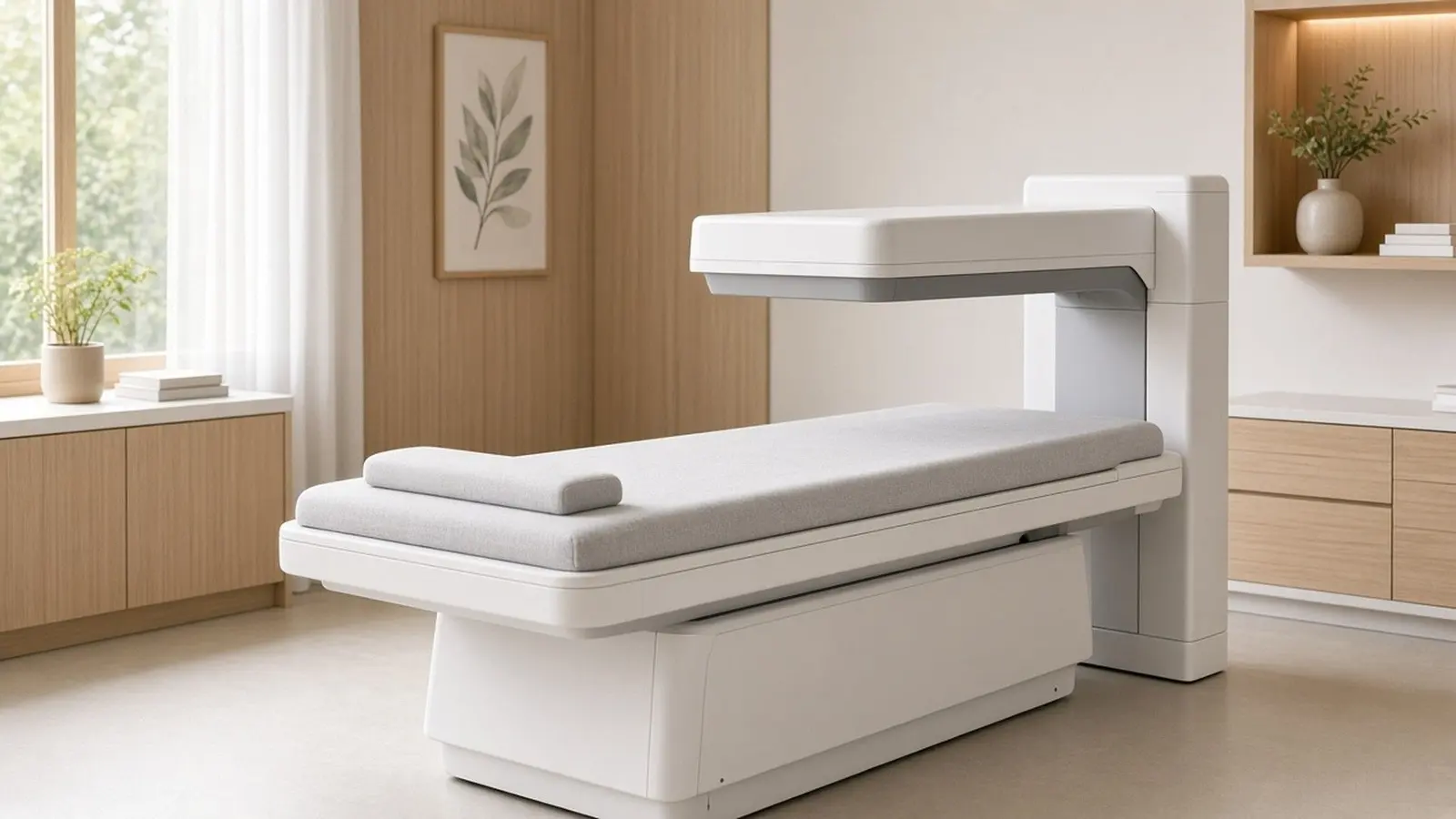
A DEXA scan measures how much calcium and other minerals are packed into your bones, usually at the hip and lower spine, and reports the result as a T-score. A T-score of -2.5 or lower means osteoporosis, -1 to -2.5 means low bone mass (osteopenia), and -1 or higher is normal. The scan takes 10 to 30 minutes and gives about 0.001 mSv of radiation, roughly three hours of natural background.
What does a DEXA scan measure?
A DEXA scan measures bone mineral density, the amount of calcium and other minerals in a defined area of bone, and from it estimates how likely that bone is to break. RadiologyInfo explains how it works: the machine sends low-dose x-rays with two energy peaks through the body, one absorbed mainly by soft tissue and the other by bone; the soft-tissue share is subtracted and what remains is the bone density. The scan is most often done on the lower spine and the hips because, as NIAMS notes, these are the bones that break most often when weak.
Two kinds of machine exist. Central DEXA is the full table scanner found in hospitals and larger diagnostic centres; NIAMS states that DEXA at the hip and spine is generally considered the most reliable way to diagnose osteoporosis and predict fracture risk. Peripheral DEXA (p-DEXA) measures the wrist, heel or finger on a small portable device. RadiologyInfo says it is used mainly for screening and is less helpful for tracking treatment, and MedlinePlus notes a low p-DEXA result usually leads to a central scan. Heel ultrasound machines seen at health camps are a third option; NIAMS states that quantitative ultrasound does not measure bone mineral density and that a positive result still needs a DEXA to diagnose osteoporosis.
RadiologyInfo lists three uses: diagnosing osteoporosis, tracking the effect of treatment, and assessing fracture risk alongside age, weight, prior fracture, family history, smoking and alcohol. See DEXA scan cost for centres.
What do the T-score and Z-score mean?
The T-score compares your bone density with a healthy young adult of the same sex at peak bone mass, and the Z-score compares it with people of your own age, sex and size. For postmenopausal women and men aged 50 or older, the T-score decides the diagnosis, using the World Health Organization cut-offs that NIAMS, MedlinePlus and RadiologyInfo all reproduce and that the 2022 Punjab DEXA study applied.
| T-score | WHO category | What it means |
|---|---|---|
| -1.0 or higher | Normal | Bone density within one standard deviation of a young adult |
| -1.0 to -2.5 | Osteopenia (low bone mass) | Below normal but not yet osteoporosis; fracture risk is raised |
| -2.5 or lower | Osteoporosis | Bone density low enough to diagnose osteoporosis |
NIAMS states that fracture risk rises by 1.5 to 2 times with each one-point drop in the T-score. The Z-score is used instead for premenopausal women, men under 50 and children. NIAMS states that a Z-score of -2.0 or less means bone mineral density is low for your age and may point to osteoporosis caused by a medicine or another disease, so a low Z-score in a younger person prompts a search for a cause such as corticosteroids or thyroid, parathyroid, liver or kidney disease.

RadiologyInfo adds a practical rule: follow-up scans should be done on the same machine at the same centre, because results from different equipment cannot be compared directly.
Who should have a bone density test?
Women aged 65 and over, younger postmenopausal women with risk factors, and anyone with a fragility fracture or a bone-thinning condition or medicine should be tested. NIAMS reports that the US Preventive Services Task Force recommends bone density testing for women over 65 and for postmenopausal women under 65 at increased risk of an osteoporosis-related fracture, and that it has made no recommendation for men. The Cleveland Clinic lists women over 65 and men over 70 as groups needing regular scans, plus adults over 50 who have broken a bone and anyone who has lost an inch and a half or more of height. The NHS advises a scan for people over 50 at risk and for younger people who smoke or have had a fracture.
RadiologyInfo lists the groups for whom testing is strongly recommended:
- Postmenopausal women not taking oestrogen, especially if tall (over 5 feet 7 inches) or thin (under 125 pounds, about 57 kg)
- A personal or maternal history of hip fracture, or smoking
- Long-term corticosteroids, some anti-seizure drugs or high-dose thyroid replacement
- Type 1 diabetes, liver or kidney disease, hyperthyroidism, hyperparathyroidism, rheumatoid arthritis or a family history of osteoporosis
- A fracture after only mild trauma, or a vertebral fracture seen on x-ray
MedlinePlus adds low body weight, lack of physical activity, long-term tobacco or alcohol use and low calcium and vitamin D intake. An orthopaedic doctor usually orders the first scan after a fracture; an endocrinologist takes over when a younger person, a man, or someone on steroids has a low result.
How do I prepare and what happens during the scan?
Stop calcium tablets 24 hours before, wear clothes without metal, and expect to lie still on a padded table for 10 to 30 minutes. RadiologyInfo and the Cleveland Clinic both say to avoid calcium supplements for at least 24 hours before the exam; MedlinePlus gives 24 to 48 hours. You can eat normally and take your other medicines. Tell the centre if you had a barium study, a contrast CT or a nuclear medicine scan recently, because RadiologyInfo advises waiting 10 to 14 days after those. Tell the technologist if you are or might be pregnant.
What happens, as MedlinePlus and RadiologyInfo describe it:
- You lie on your back on a padded table, usually in your own clothes
- A scanning arm passes slowly over the hip and lower spine while a detector moves underneath; you may be asked to hold your breath briefly
- The radiographer can stay in the room because the dose is so low, as the NHS notes
- Central DEXA takes 10 to 30 minutes; a peripheral finger or heel test takes a few minutes
- A side view of the spine to check for vertebral fractures, offered to older patients with height loss or back pain, adds a few minutes (RadiologyInfo)
MedlinePlus says results usually come within a few days. The report lists the bone density, the T-score and Z-score for each site, and often a colour chart of the ranges. RadiologyInfo cautions that spinal deformity, previous spine surgery, compression fractures or arthritis can make the spine reading less accurate, in which case the hip value or a CT measurement is used.
How much radiation does a DEXA scan give?
A DEXA scan gives about 0.001 mSv, one of the smallest doses in medical imaging. RadiologyInfo's radiation dose table lists bone densitometry at 0.001 mSv, equal to about three hours of natural background radiation, against 0.1 mSv for a chest x-ray and about 3 mSv a year from natural sources. RadiologyInfo's DEXA page puts the dose at less than one-tenth that of a standard chest x-ray and states that no radiation stays in the body afterwards. The NHS describes the scan as very safe, using a much lower level of radiation than standard x-rays, and notes that the radiographer can remain in the scanning room.
| Test | Effective dose (RadiologyInfo) | Natural background equivalent |
|---|---|---|
| DEXA bone density | 0.001 mSv | 3 hours |
| Chest x-ray | 0.1 mSv | 10 days |
| Lumbar spine x-ray | 1.4 mSv | 6 months |
The one group that still avoids the test is pregnant women. The NHS states that bone density scans and x-rays are not recommended in pregnancy, and the Cleveland Clinic says pregnant people should avoid all radiation if possible. Because the dose is so small, repeat scans are not a radiation concern; RadiologyInfo says routine re-evaluation every two years can detect significant change, and that some patients, such as those on high-dose steroids, may be rescanned at six months. MedlinePlus gives a risk-based schedule: every 2 years for high fracture risk, every 3 to 5 years for moderate risk, and every 10 to 15 years for low risk.
How do DEXA results guide treatment?
The T-score, your age and your other fracture risks together decide whether you need medicine or only lifestyle change. NIAMS states that diagnosis and treatment recommendations are based on the bone density result, age and other fracture risk factors, not on the T-score alone, and the NHS says results are used alongside a fracture risk assessment. RadiologyInfo describes a WHO online tool that combines the DEXA result with a few basic questions to predict the ten-year risk of hip and other major osteoporotic fractures.
For a normal or osteopenia result, NIAMS recommends the foundations: calcium of 1,000 mg a day for adults 19 to 50 and men to 70, and 1,200 mg for women over 50 and everyone over 70; vitamin D of 600 IU a day to age 70 and 800 IU after; strength or resistance training, which NIAMS calls the best activity for bone; no smoking and moderate alcohol; and fall prevention at home. MedlinePlus notes that a doctor may also order a calcium blood test, a vitamin D test or hormone tests to look for a cause.
For osteoporosis, or osteopenia with a high ten-year fracture risk or a previous fracture, NIAMS lists the medicine classes: bisphosphonates that slow bone loss, a RANK ligand inhibitor injection, oestrogen agonist/antagonists and hormone therapy for postmenopausal women, parathyroid hormone analogues that build bone in people at high fracture risk, and a sclerostin inhibitor for severe disease. NIAMS states that bone density testing is then used to monitor how well treatment is working.
Why does bone density matter so much in India?
Indian adults have unusually low vitamin D levels and osteoporosis appears early, so a DEXA scan is worth asking for sooner than Western age rules suggest. A 2021 systematic review and meta-analysis in BMC Public Health by Siddiqee and colleagues pooled 39 Indian studies with 38,672 participants and found vitamin D deficiency, defined as below 20 ng/mL, in 67% (95% confidence interval 61 to 73%); the South Asian figure was 68%. MedlinePlus lists the vitamin D test among the follow-up tests after a low DEXA result.
The osteoporosis burden is large. A 2022 study of postmenopausal women in Punjab, published in the International Journal of Environmental Research and Public Health, scanned 672 women aged 50 to 80 with hip and spine DEXA and found osteoporosis in 30.5% and osteopenia in 44.2%, leaving a quarter with normal bone mass. The paper cites an estimate of 61 million people with osteoporosis in India, 80% of them women, and notes that peak incidence in India occurs 10 to 20 years earlier than in Western countries.
In practice, a woman past menopause, a man over 70, anyone on long-term steroids and anyone who has broken a bone in a fall from standing height should ask for a DEXA scan rather than wait.
See a doctor the same day if a fall from standing height causes severe hip, back or wrist pain, or if you cannot bear weight; RadiologyInfo lists a fracture after mild trauma as a reason in itself for a DEXA scan. Call 112 if you cannot get up.
This guide is general information from the sources listed below, not medical advice; a registered doctor makes decisions with you.