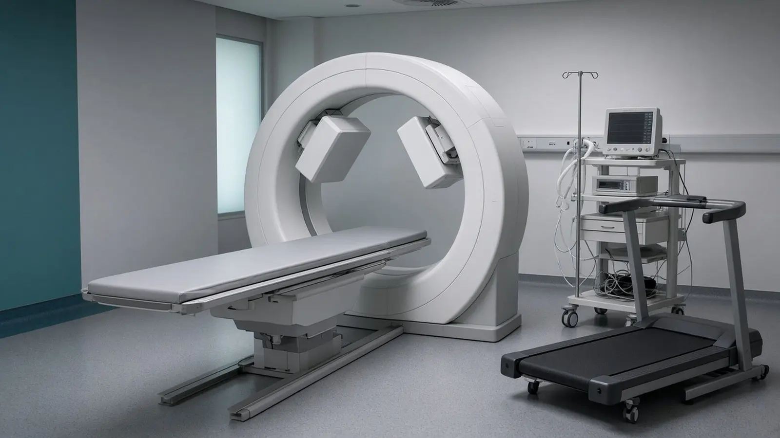
A myocardial perfusion imaging (MPI) scan, also called a nuclear stress test or stress perfusion scan, shows how well blood reaches each part of the heart muscle. A radioactive tracer is injected twice, once at rest and once when the heart is stressed by exercise or a drug, and a SPECT or PET camera photographs where the tracer goes. An area that fills at rest but not under stress points to a narrowed coronary artery; an area that never fills points to scar from an old heart attack.
What is a myocardial perfusion imaging scan?
A myocardial perfusion imaging scan is a nuclear medicine test that maps blood flow through the heart muscle. The American Heart Association (AHA) explains that it shows areas of muscle that are not getting enough blood, shows how well the muscle is pumping, and can reveal a past heart attack. It is often called a nuclear stress test; Indian prescriptions may write MPI, stress thallium or stress MIBI. The AHA notes one limit: the scan does not show the coronary arteries themselves; it shows whether, and roughly how many, arteries are blocked by the effect on the muscle they supply.
The test uses a radiotracer, a small amount of radioactive material attached to a molecule the heart muscle takes up from the blood. RadiologyInfo explains that a gamma camera detects the rays the tracer gives off, and a computer builds a three-dimensional picture; with ECG gating it also measures wall movement and overall pumping function. NIBIB describes the two camera types: in SPECT the detectors rotate around the chest to build the image from many angles, while in PET a positron-emitting tracer produces pairs of photons that travel in opposite directions and are picked up by a ring of detectors.
RadiologyInfo lists the reasons a cardiologist orders the scan: unexplained chest pain, chest pain or breathlessness on exertion, an abnormal ECG, assessing known or suspected coronary artery disease, measuring damage after a heart attack, checking a bypass or stent, and looking for heart damage from chemotherapy or radiotherapy.
Why are images taken at stress and at rest?
The two sets of images are compared because a narrowed artery often delivers enough blood when the heart is idling but not when it is working hard. RadiologyInfo describes the sequence used in most departments: an intravenous line is placed, the rest tracer is injected and images are taken within the first hour with your arms above your head for about 15 to 20 minutes. Then comes the stress phase. You walk on a treadmill or pedal a bicycle with ECG leads and a blood pressure cuff attached, and at peak effort the tracer is injected again so that it is taken up by the muscle at the moment of maximum blood flow; a second image set follows.
If you cannot exercise because of joints, balance, lung disease or a recent illness, a drug stands in for exercise. MedlinePlus names adenosine or dipyridamole, which widen the coronary arteries, and dobutamine, which makes the heart beat faster and harder. The AHA calls this a chemical or pharmacologic stress test. RadiologyInfo lists its passing effects: anxiety, dizziness, nausea, shakiness, breathlessness or mild chest discomfort.
MedlinePlus explains that after each injection you wait 15 to 45 minutes before the camera takes its pictures, and a computer then lines up the two sets. RadiologyInfo gives 15 to 30 minutes per scan and two to four hours for the whole visit, and notes that being called back for extra images does not by itself mean a problem was found.

Which tracers are used, and what is the difference between SPECT and PET?
SPECT uses technetium-99m sestamibi, technetium-99m tetrofosmin or thallium-201, while PET uses rubidium-82 or nitrogen-13 ammonia; the AHA states that the tracer is the main difference between the two kinds of MPI. A 2016 review in Acta Cardiologica Sinica by Hung and colleagues lists all five tracers with their standard protocols and effective doses, and NIBIB notes that SPECT is used primarily to diagnose and track heart disease such as blocked coronary arteries.
The two cameras differ in what they can measure. NIBIB explains that SPECT records the gamma rays from the tracer as the camera rotates, whereas PET detects the paired photons produced when a positron meets an electron. The Cleveland Clinic notes that a cardiac PET scan focuses on the metabolic function of heart tissue and can detect scar left by a heart attack, and the AHA lists PET uses that go beyond blood flow: telling living from dead muscle before deciding on angioplasty or bypass, cardiomyopathy, cardiac sarcoidosis and tumours. The 2016 review states that PET tracers can greatly reduce the radiation burden but that availability and logistics limit routine use, so most perfusion scans are SPECT.
| SPECT MPI | PET MPI | |
|---|---|---|
| Tracers | Tc-99m sestamibi, Tc-99m tetrofosmin, thallium-201 | Rubidium-82, N-13 ammonia |
| Camera | Rotating gamma camera | Ring of detectors for paired photons |
| Typical dose (2016 review) | 10.62 to 18.76 mSv | 2.96 to 7.55 mSv |
How do I prepare for the scan?
Stop caffeine, fast as instructed, bring your medicine list and inhaler, and change heart medicines only on the clinic's instruction. Every source is strict about caffeine. RadiologyInfo says to avoid coffee, tea, decaffeinated coffee, soft drinks, energy drinks, chocolate and caffeine-containing medicines, and smoking, for up to 48 hours; MedlinePlus and the AHA give 24 hours and MedlinePlus includes caffeine-free sodas and certain pain relievers.
- Food: RadiologyInfo says nothing to eat or drink after midnight; the AHA says water only for four to six hours before
- Medicines: RadiologyInfo says to ask your doctor about temporarily stopping beta-blockers or calcium channel blockers, and MedlinePlus says never stop or change a medicine without asking first
- Diabetes: RadiologyInfo and the AHA say people on insulin need specific instructions for dose and food on the day
- Erectile dysfunction tablets: MedlinePlus asks you to report sildenafil, tadalafil or vardenafil taken in the past 24 to 48 hours
- Clothing: comfortable clothes and walking shoes; no oil, lotion or cream; no jewellery
- Bring: an asthma inhaler if you use one and your prescription list
- Tell the department about a recent heart attack, heart failure, asthma or chronic lung disease, heart block, aortic stenosis or other valve disease, pregnancy or breastfeeding
The AHA says the scan may be postponed until after pregnancy if it is not urgent, and advises a nursing mother to pump enough milk for a couple of days of feeds beforehand or to use formula for one to two days afterwards, because, as RadiologyInfo states, some tracer passes into breast milk.
How much radiation does a perfusion scan give?
A standard one-day SPECT perfusion scan gives about 10 to 12 mSv, a two-day protocol up to 19 mSv, and a PET scan 3 to 8 mSv. The 2016 Acta Cardiologica Sinica review tabulates adult effective doses by tracer and protocol and states that SPECT doses generally range from about 10 to 20 mSv. RadiologyInfo's table puts natural background at about 3 mSv a year.
| Protocol | Effective dose | Source |
|---|---|---|
| Tc-99m tetrofosmin, one-day rest/stress | 10.62 mSv | 2016 review |
| Tc-99m sestamibi, one-day rest/stress | 12.10 mSv | 2016 review |
| Tc-99m sestamibi, two-day | 18.76 mSv | 2016 review |
| Rubidium-82 PET, rest/stress | 7.55 mSv | 2016 review |
| N-13 ammonia PET, rest/stress | 2.96 mSv | 2016 review |
| Coronary CT angiography | 8.7 mSv | RadiologyInfo |
| Chest x-ray | 0.1 mSv | RadiologyInfo |
Three things lower the dose. The review explains that if the stress images are normal the rest images add nothing and can be skipped, a stress-only protocol it suggests for people with a lower pre-test likelihood of disease; newer solid-state (CZT) cameras image with as little as 1 to 1.4 mSv for a single injection; and PET gives the lowest dose where available. RadiologyInfo states that nuclear medicine has been used for more than six decades with no known long-term adverse effects from such low-dose exposure, and the AHA says the body clears the tracer through the kidneys and stool, so drink plenty of water for a few days.
What do the results mean?
The report compares the stress and rest images region by region and describes each as normal, reversible or fixed. The AHA sets out the four patterns:
- Normal at both stress and rest: blood flow through the coronary arteries is normal
- Normal at rest but reduced after stress (a reversible defect): most likely a narrowing in one or more coronary arteries
- Reduced at both stress and rest (a fixed defect): not enough blood reaches that area at any time
- No tracer at all in an area: the cells there may have died in a previous heart attack, and other tests may be needed to confirm
RadiologyInfo adds that ECG-gated images report wall motion and overall pumping function. A radiologist or nuclear medicine physician reads the images and sends the report to the referring cardiologist, who explains it alongside your symptoms, ECG and risk factors.
MedlinePlus lists what follows an abnormal result: a change of medicines, coronary angiography to look at the arteries directly, angioplasty with a stent, or bypass surgery. MedlinePlus also cautions that other organs can occasionally cause a false-positive appearance, which is one reason an abnormal scan is confirmed with angiography rather than treated on its own.
Go to an emergency department or call 112 at once if you have chest pain that lasts more than a few minutes, spreads to the arm, jaw or back, or comes with sweating or breathlessness; do not wait for a scheduled scan.
What are the alternatives: stress echo, TMT and CT angiography?
A treadmill test (TMT) is the usual first test, a stress echocardiogram gives pictures without radiation, and coronary CT angiography shows the arteries themselves. MedlinePlus says echo or nuclear imaging is added when a plain exercise test is hard to interpret.
| Test | What it measures | Radiation | Where it fits |
|---|---|---|---|
| TMT / exercise ECG | ECG and blood pressure while walking on a treadmill; about 60 minutes (MedlinePlus) | None | First test for chest pain in someone who can walk |
| Stress echocardiogram | Ultrasound of wall motion before and after exercise or a drug; 30 to 40 minutes (NHS) | None (Cleveland Clinic) | Imaging without a tracer when the ECG test is inconclusive |
| Perfusion SPECT or PET | Blood flow to the muscle at stress and rest | 3 to 19 mSv (2016 review) | Abnormal or unclear ECG test, known disease, after a heart attack or bypass |
| Coronary CT angiography | Iodine-contrast CT of the arteries and plaque | 8.7 mSv (RadiologyInfo) | Low to intermediate risk chest pain, an inconclusive stress test, or new symptoms after a normal one |
RadiologyInfo lists the limits of CT angiography: heavy calcium can make the scan hard to read, an irregular heartbeat lowers image quality, contrast can worsen abnormal kidney function, and a blockage found on CT cannot be treated during the scan. A cardiologist chooses by your symptoms, ability to exercise, kidney function and local availability. This guide is general information from the sources listed below, not medical advice; a registered doctor makes decisions with you.