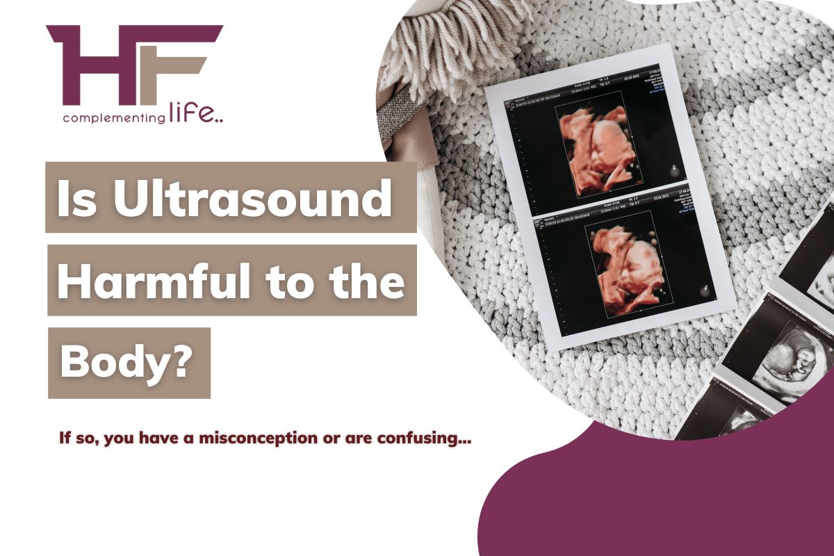
Is Ultrasound harmful to the body?
What do you think, a test that involves the diagnosis of an unborn baby, can be harmful? If so, you have a misconception or are confusing an ultrasound with some other diagnostic methods.
CContents
Is Ultrasound harmful to the body?
No doubt, most of the diagnostic methods, be it an X-ray, a CT Scan, or an MRI, can create Sid effects and lead to complications in the near future if not executed properly. It is because of the exposure to ionizing radiation amidst the diagnosis. Herein, an extra dosage of the radiation is enough to fall heavy on your health condition.
However, not in the case of an ultrasound!
Are you wondering why so? Well, it is simply because the process of ultrasound does not encompass any ionizing radiation. It is totally different from the other diagnostic procedures.
Ultrasound testing, emerging into the picture of medical technologies in 1956 through the research and development of Ian Donald in Glasgow, is indeed a safe process and bears no harmful effect on the human body. Ultrasound is quick & painless, 99% devoid of side effects or further complications. That is precisely why it is considered the most common & significant imaging test to detect a vast range of deficits & diseases.
But,
What is the proof of pedigree?
Yes, that is exactly why we are here today, writing this note to you all, offering the proof of pedigree you are looking for!
This article on – Is Ultrasound harmful to the body – aims to give you a thorough insight on the ultrasound procedure and its difference from other imaging tests. It shall also entail a brief history of the ultrasound method, its development and different approaches, what organs it can show, and what disease it can examine. In fact, the article shall also tell you when your doctor may recommend you to have an ultrasound!
Are you ready? Then, Gear up to learn it all!
History of Ultrasound Test & its Development through Time: A Note!
Although the practice of ultrasound began after the second world war, essentially from Glasgow, the concept of visualizing internal body organs through sound waves began even before that, right from 1908. In fact, the modality of ultrasound imaging took the stage in the late 1700s as a part of a theoretic explanation for bat aviation.
From 1729 to 1799, Lazzaro Spallanzani, a priest & Italian physiologist, carried out a set of experiments to demonstrate how & why bats have the ability to fly at night time. It was these experiments that made him realize that a bat if made blind, can still fly through space, but if it is deaf, even in one particular ear, it cannot. Hence, the basis of navigation via sound stood before the human eye!
Subsequently, in 1826, Swiss physician Jean-Daniel Colladon laid out that the speed of sound is faster than in the air. Then, in 1908, Karl Dussik, an Austrian Neurologist, took an attempt in using ultrasound to depict the transformation of brain ventricle size as a part of tumor growth. During this time, the wand-like instrument, called a transducer, used in the process of diagnosis lay placed on both sides of the patient’s head.
In 1928, SY Sokolov, a physicist from the Soviet Union, gave a proposal of using ultrasound to detect flaws in metallic structures. At that time, ultrasound was popular for industrial use. In 1949, George Ludwig from the US Naval Military Research Institute was performing research on gallstones that lay embedded in the soft tissues. His research comprised the use of transmission techniques. It is this very instance when he pioneered the assessment of interactions between animal tissues & high-frequency sound waves.
The successful use of ultrasound in clinical practices only started in 1956, when Ian Donald sought to measure the parietal diameter of a fetal head by using a one-dimensional amplitude mode transducer. After two years, ultrasound was again used to scan a female genital tumor. Later on, Brown invented a two-dimensional scanner to visualize the density of tissues. Perhaps, the turning point in the application of ultrasound in medical testing!
Ultrasound, the process of using sound waves to diagnose the inside of your body, requires only three pieces of equipment, namely, the probe or transducer, a computer console, and a video monitor. Today, it is the most versatile technique to diagnose internal health conditions and holds various methods or practices. While the traditional ultrasound failed to detect as deep as your blood flow, development took place. Thereupon, came into the picture Doppler Ultrasound. Doppler Ultrasound is the process in which your bloodstream lay diagnosed with almost 78% sensitivity. Yet when this failed to examine the direction of blood flow, the color doppler approach came to highlight the direction of blood flow within your veins & arteries.
With the advancement of medical science, another ultrasound approach came into existence, and it is known as the endoscopic ultrasound. In this process, the ultrasound technique works in combination with the endoscopic techniques, creating images more precise than before! Endoscopy, which developed in 1911, when got collaborated with ultrasound imaging, gave way to a new dimension in the field of imaging tests. Initially, this process was meant to improve the screening of the pancreaticobiliary system, i.e., obstruction or blockages, injury, or tumors in the pancreas & bile ducts. Later, this method became significant in screening the gastrointestinal tract and nearby areas as well!
Further development in medical technologies led to another advanced ultrasound approach called Saline Infusion Sonography. This particular method, where a small amount of salt solution stands requisite to create a clear visualization of the internal structures, especially of the endometrial cavity, stood popular since the first decade of 2000 in the field of obstetrics & gynecology. It is because this ultrasound technique helps diagnose abnormalities like endometriosis and also serves in assessing the uterus shape & size during an ovarian treatment therapy.
Today, ultrasound is the most common & reliable diagnostic modality across the world and specifically serves to monitor the condition of pregnancy on a routine basis. So, do you think that a medical practice that came up all the way, inhabiting transformations & development throughout, can be harmful to your body? Perhaps, not!
It is more because the people at work are expert medical professionals! They are the ones who have learned & practiced ultrasound techniques for years and thereafter worked as a team! The team of radiology!
Do you want to know who they are? Then, do not hesitate to read below!
Who conducts an Ultrasound & Where?
Well, when you opt for an ultrasound testing, you must know that there is no one single person or professional to carry out the examination but a team of medical caregivers that includes –
1. A Radiologist. The one, who supervises the entire diagnostic process, be it for an ultrasound or other medical tests like blood tests, MRIs, endoscopy, colonoscopy, and so forth. It means the radiologist is the one leading the team and shall instruct the other members to conduct the test with dexterity & precision.
2. An Ultrasound Technician, who is also referred to as a Sonographer, is the second person on the team. The sonographer is the main man or woman at work. In other words, the sonographer is the one who shall conduct the test, from introducing the transducer to your body, inserting it into your vagina, or pressing it over your skin, & continuing to do so until images from multiple angles are taken.
3. Medical Staff, who works as a helping hand of the sonographer. This person shall be by your side the entire time and assist you in case you need something or want to say something. The medical staff shall also aid you if and when you feel any discomfort and shall take you to the waiting room after the test stands complete.
Yes, three members work as a team to help assess your internal abnormalities on an outpatient basis. It means that you do not need to stay back for long and can return to your normal schedule once the test is over.
On this note, let me tell you that ultrasound can take place in a hospital, a clinic, or an exclusive diagnostic center, depending on the recommendation of your doctor. If you are selecting the center yourself, make sure you choose the one which has all state-of-the-art equipment and the best medical professionals in place! That is when you can go ahead to get your ultrasound done with a deep sigh of relief!
Herein, an immediate question is likely to arise! What is the procedure? Or, in other words, how is the test done?
Wait!
I will tell you how, but for that, you have to read the next segment, word for word, and prepare yourself accordingly!
How is an Ultrasound Performed?
By now, you must be aware of the fact that an ultrasound stand performed using high-frequency sound waves. These sound waves appear from the transducer and reflect back to the transducer again on encountering any organ or tissue. That is how the transducer can convert the echoes into electric signals and send the images of your internal organs & tissues right on the monitor screen.
This process shall take a maximum of 30 minutes, but usually, it wraps up within 15 to 20 minutes!
Do you want to have a step-by-step guide to know what happens in that 15 minutes? Well, see-through!
1. When you enter the test lab, you shall come across different state-of-the-art equipment lined up one after the other. You may be mesmerized to see the various technologies, but please do not halt. Can you see a large examination table or bed therein? That is your destination! That is where you have to lie down for the ultrasound test.
2. Before entering the hall, the medical staff may give you a gown to wear. But, sometimes, they may not! For this reason, it is advisable to wear a loose dress on the day of the test and avoid all kinds of accessories. Even if you wear any accessories, make sure you remove them before the test starts, as any kind of metallic objects or accessories may interrupt the diagnosis and tamper with the test results. Now, you have to slip off your dress & expose the definite body part for the examination.
3. Once you do so, the sonographer shall begin his work on the instructions of the radiologist. He shall take out the transducer and apply a lubricant over it. This lubricant is meant to serve in two ways. First, it eliminates air from the space between your skin and the transducer, and second, it wipes out all kinds of pain and discomfort when the transducer is inserted into your vagina or pressed over your skin.
4. In the case of a vaginal ultrasound where the transducer gets inserted into your pelvic region through the vaginal opening, there is an additional step. The sonographer, after applying the gel, shall also cover the tip of it with a plastic sheet. This sheet appears somewhat like a condom and helps reduce painful sensations if any.
5. The next step is simple! The sonographer shall ask you to stay still for a while with your knees bent and feet at a stir-up position, and then either insert the transducer through your vagina or mouth or press & move it over your skin near the area that needs to be assessed. In doing so, sound waves originating from the transducer tend to travel through your body.
6. That is when they come across your organs, tissues, arteries & veins. Thereupon, reflecting back to the transducer! Of course, you cannot hear the sound. Nor can you feel it passing through! But, when the faint echoes culminate into electric signals, the imaging pops up on the computer screen, and the radiologist can see the condition of your organs & tissues.
7. When one imaging does not suffice, and the radiologist cannot detect the problem, he shall instruct the sonographer to retake the process of pressing the transducer again from a different position. At that time, you shall have to shift to the said position and hold on to that until the second imaging shows up again. This particular step shall give the radiologist an opportunity to extract ultrasound imaging from various angles. Thereafter, aid your doctor in precisely detecting the defect!
8. That’s all! Your doctor shall now instruct the sonographer to remove the transducer. Now, you shall be free to leave the test lab and move to the waiting room. Here, you can use the washroom and dress up as you came. Meanwhile, the medical experts shall keep an eye on you to confirm that no side effects appear. Once they are assured, you can drive back home or to your office. However, it is always better to get someone along with you, nothing but your support. The one, who can help you reach your destined location after the test!
Do not worry about the report. The medical team shall prepare & send it straightaway to your doctor or send it to you via an online medium. In some situations, you may have to travel back to the center to collect the report and if you want to avoid this step, make sure you book your diagnosis via www.healthfinder.com.
Ultrasound Vs Other Imaging Test: What are the differences?
Are you not sure about the benefits of this test? Are you looking forward to knowing how this test is better than so many other imaging tests that are undertaken these days?
Well, let me tell you that the ultrasound imaging test is one of the most reliable tests today. It is because the process leaves room for no disadvantages at all!
How?
Discover right now!
1. Difference between Ultrasound & Endoscopy and/or Colonoscopy –
Both the process of endoscopy and colonoscopy involves an invasion into your body through the introduction of a small slim tube-like instrument with a camera and light attached to it, called the endoscope or a similar instrument known as a colonoscope through your mouth, rectum, or vaginal opening. So, if mishandled can lead to bleeding. On the contrary, an ultrasound is either non-invasive or minimally invasive, and your body shall have no wear & tear. Perhaps, it is one of the most significant markers that makes ultrasound different from endoscopy and colonoscopy!
Another is the non-exposure to ionizing radiation, and therefore, non-vulnerable to threats like cancer, unlike an endoscopy or colonoscopy. In fact, the use of sedatives is very rare and mostly not present in traditional ultrasound. However, endoscopic or colonoscopic testing always holds the use of sedatives that may give rise to side effects like vomiting, dizziness, rashes & infections, fever, abdominal pain, and sometimes complicated conditions like kidney damage.
2. Difference between Ultrasound & X-Ray and/or CT Scan –
Even though both the methods, X-Ray and CT Scan, are non-invasive like ultrasound, they incorporate some amount of radiation dosage while diagnosing your internal structures. Such dosage is non-existent in the practice of ultrasound. Therefore, an ultrasound is not prone to complications like cardiovascular diseases, inflammation of organs, tumors, polyps, cancerous growth, and so forth that may arise after an X-ray or CT Scan.
On the same note, differences also lie in terms of diagnostic efficiency. While an X-ray or CT scan is more significant in assessing bones and gas-filled organs, an ultrasound screens much deeper into the internal details of an organ or tissue. Besides, X-Rays and CT scans are typically more expensive than ultrasound. In India, an ultrasound may cost up to a maximum of Rs. 1500, but an X-Ray or CT scan is likely to go as high as Rs. 3200.
3. Difference between Ultrasound & MRI –
MRI, or magnetic resonance imaging, is the process that relies on magnets for the production of 3D images of your internal body. But, an ultrasound, as you know, uses high-frequency sound waves instead of any magnetic resonance. On the same note, an ultrasound is more widely accessible because the process is much quicker than an MRI, which may even take up to 2 hours.
Well, an MRI involves the combination of magnetic fields & radio waves, and we ought not to confuse that these radio waves are ionizing radiation. It means there is an absence of ionizing radiation in both ultrasound and MRI testing, and so are serious threats like cancer or cell damage. However, in comparison to an ultrasound that can be performed on any human body, irrespective of the present medical conditions the individual holds, MRI lacks such a capability. People with an implanted pacemaker, bone-growth stimulators, neurostimulators, prosthetic devices, intrauterine contraceptive devices, or any iron-based metallic implants cannot undergo an MRI due to the presence of a strong magnetic field in it.
Now that you already know how ultrasound stands out over other imaging tests, you must be keen to learn which organs can this widely accessible & versatile diagnostic method show. Let us wait no time and quickly move into the details!
Which organs can an Ultrasound show?
An ultrasound, being various in technique, depends upon the location of the examination and can detect a wide range of organs. Thus, for learning which organs an ultrasound can show, you have to simultaneously learn the definite type of ultrasound in practice!
1. Breast or Chest Ultrasound – As the name suggests, the ultrasound method in which your breast and nearby area lay diagnosed is called a breast ultrasound. So, the organs that show up are your mammary glands, lungs, breast bones or ribcage, cardiovascular organs, and so forth.
2. Pelvic & Abdominal Ultrasound – This method of ultrasound can be categorized into three types, namely, 1) vaginal ultrasound, 2) abdominal ultrasound, & 3) rectal ultrasound. They are the ones that examine your pelvic & abdominal organs, such as the placenta, uterus, ureter, cervix, urethra, fallopian tube, rectum, ovaries, urinary bladder, kidney, gallbladder, appendix, pancreas, liver, stomach, bile ducts, small intestine, large intestine, spleen, colon, etc.
3. Carotid Ultrasound – Carotid Ultrasound is the physical examination that covers the screening of carotid arteries, i.e., pair of blood vessels located on your neck.
4. Thyroid Ultrasound – Likewise, thyroid ultrasound entails the diagnosis of your thyroid gland and other organs of the endocrine system like the pituitary gland, hypothalamus, parathyroid, adrenal glands, etc.
5. Musculoskeletal Ultrasound – Above all, the musculoskeletal ultrasound is meant to examine your muscles, joints, tendons, ligaments, & nerves underlying throughout your body.
Thus, whatsoever abnormalities pop up in the aforementioned organs, an ultrasound test shall be efficient enough to detect!
Which medical conditions can an ultrasound assess?
Undoubtedly, a versatile imaging test like ultrasound can trace down an unending list of medical conditions within a human body! It is because of the details it peeps into through the sound waves that tend to travel through your body to various corners, within your tissues, bones, & muscles.
Do you want to glance through the various abnormalities that ultrasound can trace? Get ready because here they go!
- Cysts, Fibroids, Polyps, or benign tumors.
- Urinary Tract infections.
- Infections in your Liver, Pancreas, Stomach, Bladder, Kidney, Ureter, Uterus, and so forth.
- Pelvic Organs Prolapse, like your womb hanging down into your vagina or maybe your bladder bulging into the anterior vaginal walls.
- Pelvic inflammatory diseases occur from sexually transmitted infections.
- Pelvic floor dysfunction, i.e., the condition in which your pelvic floor muscles fail to relax and coordinate.
- Kidney Stones, Gallstones, Bladder Stones, etc.
- Fatty Liver Disease.
- Hepatitis, or in other words, inflammation of your liver.
- Kidney Blockage or injury.
- Bladder Dysfunction or urinary incontinence.
- Overactive bladder.
- Bladder inflammation or cystitis.
- Interstitial cystitis.
- Pocket-like structures that grow along your urethra are called Urethral Diverticulum.
- Enlarged spleen and spleen injury.
- Inflammation of the pancreas or pancreatitis.
- Chronic pancreatitis, is also called cystic fibrosis.
- Diverticulitis or Inflammation of the colon.
- Colon Cancer.
- Appendix inflammation or appendicitis.
- Inflammation of the stomach lining or gastritis.
- Polycystic Ovary Syndrome (PCOS).
- Hernia, Peptic Ulcers, and Celiac Disease.
- Gastroesophageal Reflux Disease (GERD).
- Cancer in the Kidneys, Liver, Uterus, Stomach, or Intestines.
- Ovary Torsion, Endometriosis, Vaginal blockage.
- Fecal incontinence or bowel control problem,
- Pelvic congestion syndrome.
- Irritable bowel syndrome.
- Anal Fissure.
- Colitis.
- Hemorrhoids.
- Breast Cancer, Lung Cancer, and so forth.
- Lumps in your breasts.
- Fibrocystic Breast Disease.
- The condition where your breast tissues get injured due to excess fat is referred to as Fat necrosis.
- The medical conditions in which excess male breast tissue tends to develop. This particular condition is known as Gynecomastia.
- Fluid Collection in the abdominal cavity.
- Narrowing or thickening of the stomach muscles.
- Pneumonia, Bronchitis, & Pulmonary Fibrosis.
- Chronic Obstructive Pulmonary Disease (COPD).
- Coronary Heart Disease.
- Peripheral Artery Disease.
- Aortic Aneurysm, or in other words, a balloon-like bulge in your aorta.
- Goiters in the Thyroid Gland.
- Overactive or Underactive Thyroid.
- Plague build-up in the Carotid Arteries.
- Tendonitis or Inflammation of a tendon.
- Carpal Tunnel Syndrome where the median nerve stands compressed, largely.
- Bursitis, i.e., the condition where the bursae or fluid-filled sacs cushioning the bones, tendons, & muscles tends to be affected or damaged.
- Ligament sprains, muscle strains, & joint effusion.
- Fistulas and many more!
This the where the most crucial questions appear!
How would your doctor understand that you may be affected by either of these medical conditions?
Of course, she must be predicting the abnormality based on something!
But what is it?
Well, they are nothing but the symptoms you have!
When may your doctor recommend an ultrasound?
So, your doctor may recommend an ultrasound when she predicts the plausible cause behind the different physical problems you have in your daily life.
Tell us,
Which among the following are you witnessing for the last few days?
- Persistent pelvic pain, abdominal pain, or backaches, accompanied by fever.
- Muscles Cramps that get frequent at night.
- Blood in the stools or urine.
- A burning sensation or sudden discomfort while urinating.
- Frequent urination.
- Fatigue, dizziness, & nausea.
- Gas or Bloating.
- Unintended weight loss, and that too abruptly within a few days or weeks.
- Chest pain & Chronic heartburn.
- A Tenderness near your abdomen, stomach, or pelvic muscle floors.
- A tenderness near your nipples, neck, or chest.
- Missed, Heavy, or prolonged periods.
- Abnormal vaginal bleeding or spotting in between two menstrual cycles.
- Painful & burning sensation while having sexual intercourse or an Inability to have an orgasm.
- An unusual vaginal discharge comes together with an unpleasant odor.
- Urine leakage while you cough, walk, laugh, or sneeze.
- Abnormal hair growth in the areas near your chest, stomach, or back.
- Trouble breathing or severe breathlessness at frequent intervals.
- Change in breast size, shape, & contour.
- Rashes & Redness near your nipples.
- Unusual nipple discharge accompanied by breast pain.
- Irritation or itchiness in your breast skin.
- Swelling or lump-like sensation near your nipples.
- Difficulty in swallowing food, vomiting, sore throat.
- Constipation or Diarrhea.
- High or low Blood Pressure.
- Persistent indigestion.
- Pain in your neck, throat, or jaw.
- Pain in the rectum or anus.
- Vagina bulging out, or so is the anus.
- Inability to conceive for long
- Repeated abortion or miscarriage.
- Multiple sites of pain, specifically near the limbs.
- Inability to move your joints suddenly.
- Severe headache alongside a loss of balance.
- A sudden loss of vision and difficulty speaking or forming words.
And myriads of others! Whenever you encounter any of these, it is, therefore, an advisory to call your doctor and discuss it with her.
Summing Up:
Thank you for reading the note till the end. Thy is the sum up, and we shall come back again with another article on ultrasound, thereby helping you go through the process smoothly! Meanwhile, if you want to book an ultrasound, you can simply go to our website and ask for the quotes. HealthFinder offers flexible booking schedules & enormous discounts together at a time. It means you neither have to wait in a queue to get your ultrasound done nor do you have to pay any extra money for a fast process.
Hurry up! We are waiting to serve you!


Subscribe
0 Comments

Comments